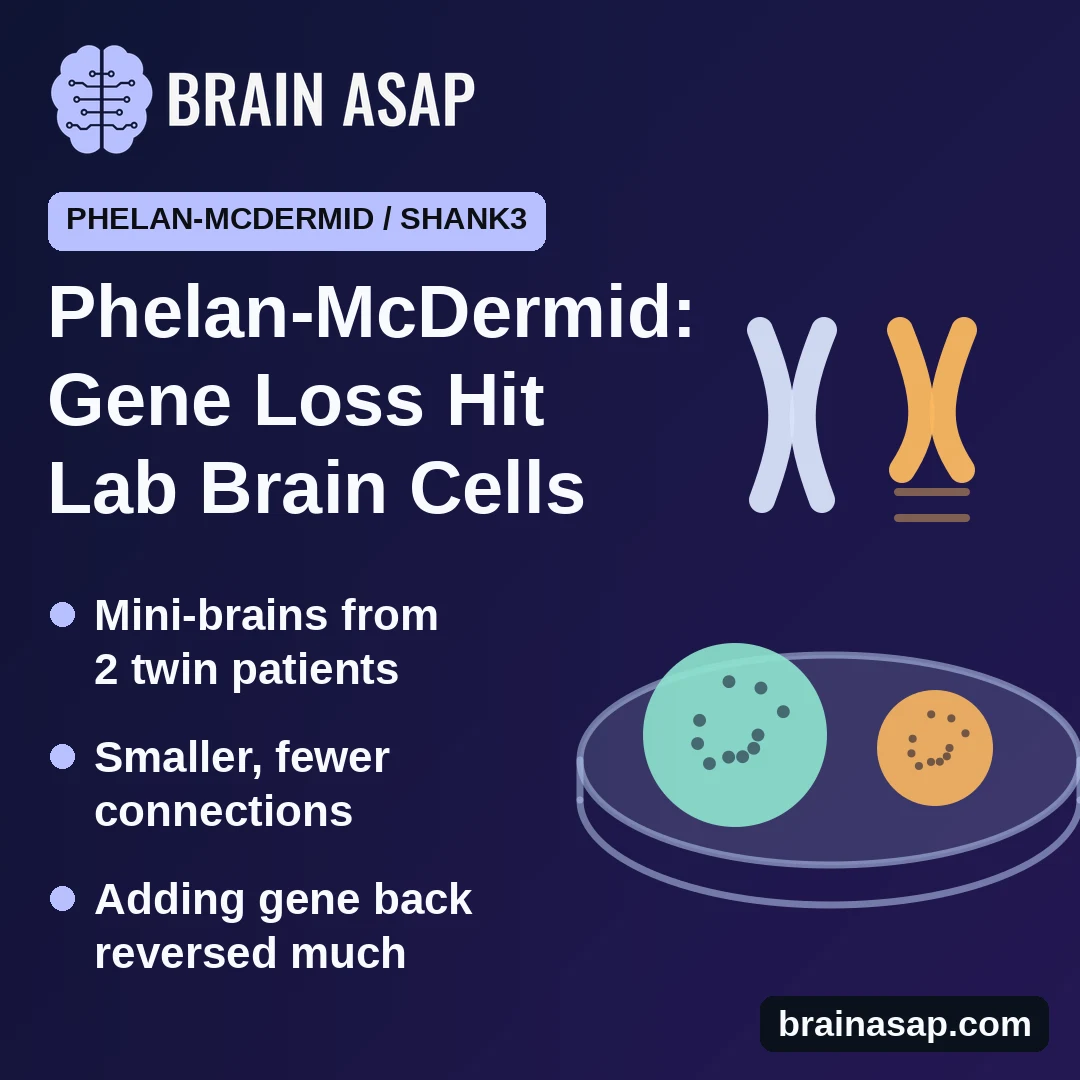
TL;DR: Researchers turned cells from twins with Phelan-McDermid syndrome, a rare genetic condition that causes intellectual disability and often autism, into lab-grown clumps of brain tissue. The organoids were smaller than those from control lines, and the patient-derived neurons had a lower density of connections. Raising SHANK3 expression largely reversed the differences in the lab.
Key Findings
- Smaller tissue clumps: patient-derived clumps ended up smaller, which the researchers traced to stem cells in them dividing less.
- Wrong mix of cells: fewer of the stem cells became signal-sending neurons; more became support cells and signal-damping neurons.
- Simpler, quieter neurons: the neurons that did form had less branching, fewer connection points and less electrical activity.
- Putting the gene back helped: delivering extra copies of SHANK3 largely undid these differences in the lab dish.
Source: Neuroscience Letters (2026) | Published abstract only
Phelan-McDermid syndrome comes from losing a piece of the end of chromosome 22. Children with it typically have intellectual disability, very delayed or absent speech, weak muscle tone, and in many cases an autism diagnosis.
Attention has long focused on a single gene inside that lost stretch, SHANK3, which builds scaffolding at the synapse, the junction where one neuron passes a signal to the next. What is less clear is whether the gene also matters much earlier, while the brain is still being built.
Growing Brain Tissue From Two Patients
A 2026 study in Neuroscience Letters took cells from twins with the syndrome, both missing a 1.7 million-letter stretch of chromosome 22 that includes SHANK3.
Those cells were reprogrammed into stem cells able to become any cell type, then grown 2 ways:
- Flat cultures: neurons grown in a single layer in a dish, easy to measure one cell at a time
- Organoids: 3-dimensional clumps, sometimes called mini-brains, that self-organize into something loosely resembling early brain tissue
What Went Differently
Organoids carrying the deletion were smaller. According to the published abstract, this came down to the neural progenitor cells, the dividing stem cells that supply a growing brain with new neurons, multiplying less than they should.
Those progenitors also produced a different mix of cells. Fewer became excitatory neurons, the ones that pass signals on. More became astroglia, the brain's support cells, and GABAergic neurons, which damp signals down.
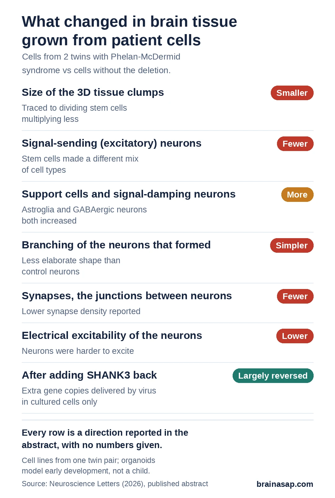
Neurons themselves were simpler in shape, carried fewer synapses and were harder to excite electrically.
Adding the Gene Back
To test whether SHANK3 was really behind all this, researchers used a virus to deliver extra copies of the gene into the patient-derived cells.
Raising SHANK3 expression largely reversed the differences. The result supports a contribution from SHANK3 in these cells, which also lacked other genes in the deleted region.
What a Dish Cannot Show
- Read from the abstract only: the published abstract gives the direction of each finding. It does not give the number of organoids or cells analyzed or the size of the effects
- Two patients, and they are twins: the cells come from a single family and a single deletion, so this is not a survey of the syndrome
- A deletion, not just one gene: that 1.7 million-letter stretch removes other genes too, though the rescue experiment points at SHANK3
- Organoids are not brains: they have no blood supply, no sensory input and no behavior, and they model early development rather than a living child
- Gene delivery in a dish is not gene therapy: adding SHANK3 to cultured cells says nothing about safety or benefit in a person
Why the Timing Matters
The findings suggest SHANK3 plays a role while the cells that make neurons are still multiplying, before those neurons form synapses.
Whether these dish-grown findings hold in larger sets of patient lines, and in tissue further along in development, is what the next studies will have to establish.
Citation: DOI: 10.1016/j.neulet.2026.138783. SHANK3 drives abnormal neurodevelopment in iPSC-derived 2D and 3D brain models of Phelan-McDermid Syndrome. Neuroscience Letters. 2026. PMID: 42998816.
Study Design: Laboratory study of induced pluripotent stem cell lines from 2 twin patients with a 1.7 Mb 22q13 deletion including SHANK3, differentiated into 2-dimensional neuronal cultures and 3-dimensional cerebral organoids, with lentiviral SHANK3 overexpression as a rescue experiment.
Sample Size: Cell lines from 2 patients (twins); the abstract does not state the number of control lines, organoid batches or cells analyzed.
Key Statistic: None available. The published abstract reports reduced organoid volume, impaired neural progenitor proliferation, decreased excitatory neurons with increased GFAP-positive astroglia and GABAergic neurons, simplified neuronal morphology, reduced synapse density and diminished excitability, all largely rescued by SHANK3 overexpression, without numerical values.
Caveat: This article is based on the published abstract alone; the full text was not available. Cell lines from a single twin pair; organoid and culture models cannot show behavior, and the rescue was performed in cultured cells, not in people.