TL;DR: In a German study of 34 people recovering from LGI1 encephalitis, a rare autoimmune brain disease, brain scans showed changes well beyond the memory centers usually blamed. People with more of these changes had poorer thinking scores and recovered less.
Key Findings
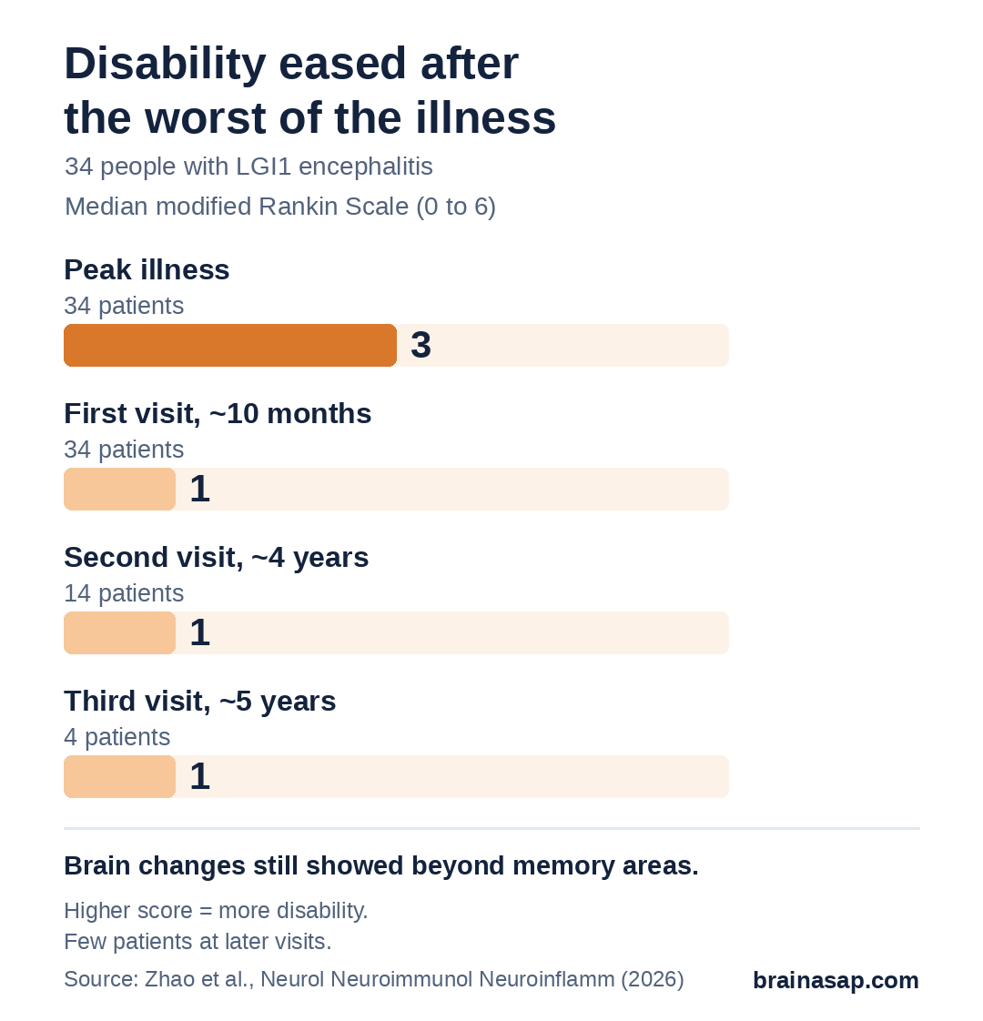
- Most improved: Median disability score fell from 3 at the worst point to 1 by the first study visit.
- Changes beyond memory areas: Frontal, temporal and parietal cortex and deep brain structures differed from healthy people.
- Linked to thinking: More changes went with poorer overall thinking scores.
- Relapses and time: Longer illness and relapses were linked to further loss in deep brain areas.
- New measure helped: A brain-shape complexity score picked up changes that volume missed.
Source: Neurology: Neuroimmunology & Neuroinflammation (2026) | Zhao et al.
In LGI1 encephalitis, the immune system makes antibodies against a protein that helps nerve cells signal. It often starts in older adults with brief jerking seizures of the face and arm, memory loss and confusion. Many people improve with treatment, but some are left with lasting thinking problems.
Doctors have long treated it as a disease of the memory centers deep in the temporal lobes. A team at Charité in Berlin looked across the whole brain to see whether that picture is too narrow.
34 Patients Followed for Up to 5 Years
The study included 34 people with LGI1 encephalitis (median age 67, 29% women) and 34 healthy people matched for age and sex. Patients had MRI scans, thinking tests and disability ratings at a first visit about 10 months after symptoms began. Fourteen returned at about 4 years and four at about 5 years.
Besides measuring the size of brain regions, the researchers used fractal dimension, a score of how complex and folded a brain region’s shape is. A lower score suggests a simpler, more worn-down structure, even when size has not changed much.
Most Patients Recovered a Lot
At the worst point of illness, the median score on the modified Rankin Scale, a 0 to 6 disability scale, was 3, meaning moderate disability needing some help. By the first study visit it was 1, meaning symptoms without significant disability, and it stayed there at later visits. Eight patients (24%) had a relapse, usually within the first year.
Brain Changes Reached Far Beyond the Memory Centers
Compared with healthy people, patients had lower shape-complexity scores across a band of frontal, temporal and parietal cortex, including language areas. Deep structures, the hippocampus and the nucleus accumbens, were both smaller and less complex. In the outer cortex, only the complexity score showed differences; plain volume did not.
These changes mattered. Lower complexity in the pallidum, a deep movement and motivation hub, went with less recovery on the disability scale. A smaller nucleus accumbens went with less recovery on an encephalitis symptom scale. Across 31 brain regions, more changes were linked to poorer overall thinking scores, and the complexity score was the stronger marker in 19 of them.
Relapses and Longer Illness Tracked With More Loss
Over time, longer illness was linked to further shrinking and simplifying of deep structures such as the thalamus, caudate and putamen. Relapses were linked to more loss in the nucleus accumbens and caudate. Older age at onset was linked to changes across the brain.
Small Numbers at Later Visits
- Few follow-ups: Only 14 patients had a second scan and 4 a third.
- Referral clinic: Patients entered at different times after onset.
- No control follow-up: Healthy people were scanned once, so normal aging over time could not be compared.
- Thinking links unadjusted: The authors say age and education need fuller adjustment.
- Observational: Cannot show the brain changes caused poorer recovery.
A Whole-Brain Disease Needs Whole-Brain Follow-Up
For patients and families, the study supports a broader view of LGI1 encephalitis: recovery can be good, but the disease leaves traces across the brain, and relapses appear to add to them. That is one more reason to catch and treat relapses quickly.
The open question is whether early, stronger immune treatment can prevent these widespread brain changes, which larger studies with scans from the first weeks of illness could answer.
Citation: DOI: 10.1212/NXI.0000000000200649. Zhao W, Wurdack K, Bartels F, et al. Structural brain changes are linked to clinical and cognitive outcomes in anti-LGI1 encephalitis. Neurol Neuroimmunol Neuroinflamm. 2026;13(6):e200649.
Study Design: Observational cross-sectional and longitudinal MRI study at a single referral center.
Sample Size: 34 patients (52 visits) and 34 age- and sex-matched healthy controls.
Key Statistic: Median mRS 3 at peak illness vs 1 at first visit (p < 0.001); pallidum shape complexity linked to recovery (ρ = 0.51, FDR-corrected p = 0.022).
Caveat: Small follow-up subgroups; no longitudinal control scans; observational design.