TL;DR: In a 2026 study, a self-teaching computer program studied microscope images of rat brain slices, stained to show the fatty insulation around nerve fibers, with no labels saying what anything was. It sorted the tissue into real brain regions on its own and flagged a pattern that covered far more of the brain in a rat with an obvious mild brain injury, though only 4 rats were tested.
Key Findings
- Learned without labels: the AI, a type called an autoencoder, was trained on 420 brain slices from 9 rats with no human notes on what the tissue was.
- Found brain regions on its own: it grouped tissue into white matter of different densities and gray-matter regions such as cortex layers and parts of the hippocampus.
- Flagged an injury-linked pattern: one tissue group covered about 5% to 6% of the displayed brain tissue in uninjured rats, 8.6% in a rat with subtle injury and 38.8% in a rat with obvious injury.
- Less intact white matter after injury: about 13% in uninjured rats vs. 9.7% in the more injured rat, mostly on the injured side.
- Small edge over simpler methods: the AI kept fine nerve-fiber detail slightly better than a standard math method and a general-purpose image AI.
Source: Brain Structure and Function (2026) | Estela et al.
Studying brain tissue under a microscope still often means an expert looking at slide after slide and outlining regions by hand. It’s slow, and different experts can draw different lines.
A 2026 study in Brain Structure and Function, by researchers at the University of Eastern Finland, asked whether an AI program could learn what brain tissue looks like with no human labels at all, and then notice when something was wrong.
Teaching an AI With No Answer Key
The images came from an earlier experiment on 13 adult male rats. Seven had a mild brain injury, made by a quick pulse of fluid pressure on the left side of the brain. Six had the same surgery without the injury.
Brain slices were stained to show myelin, the fatty coating that insulates nerve fibers. Each slice image was cut into thousands of small square patches.
An autoencoder is an AI that learns by squeezing each patch down to a short summary of 256 numbers and then trying to rebuild the original image from it. To do that well, it has to learn which features matter. Nobody told it what white matter or injury looks like.
Training used 420 slices from 9 rats. Testing used 194 slices from 4 different rats: 2 uninjured and 2 injured.
Sorting Tissue Into Brain Regions
A separate step then sorted the patches into groups based on those learned summaries. When the groups were painted back onto the slices, they lined up with real anatomy:
- White matter split by how densely packed the nerve fibers were
- Layers of the cortex
- Subregions of the hippocampus, a memory center
Compared with a standard math method (principal component analysis) and a large general-purpose image AI (DINOv3), the autoencoder separated cortex layers more clearly and kept groupings more consistent.
On rebuilding images, the differences were small. Principal component analysis matched pixel brightness a bit better, while the autoencoder kept fine nerve-fiber structure a bit better.
Did the AI Notice the Injury?
The result? In the more injured rat, clearly. Tissue from the 4 test rats was sorted into 9 groups, and the team checked how much of each displayed brain section fell into each one.
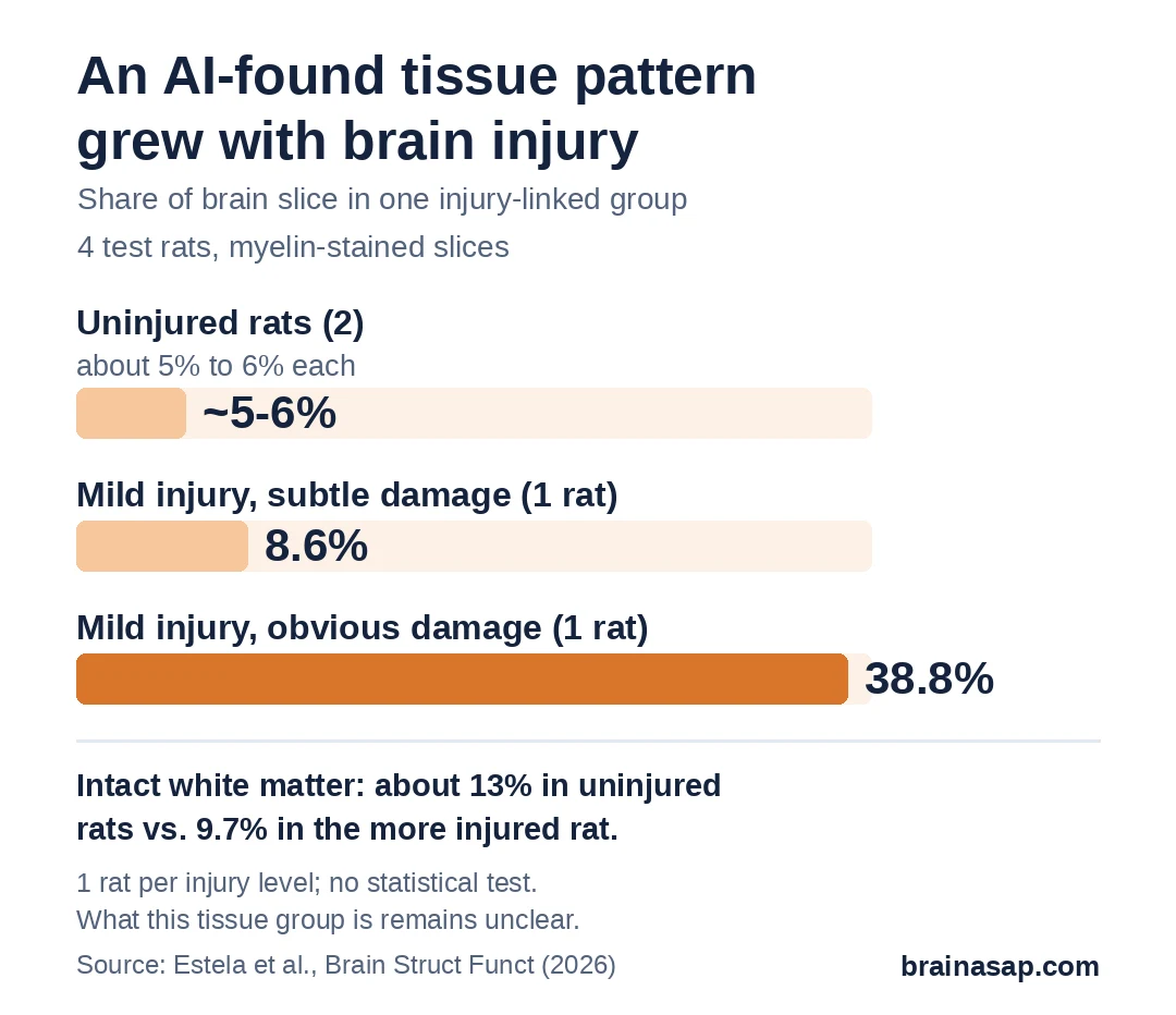
One group, mostly tissue at the edges of brain structures and in the hippocampus, stood out. It barely registered in uninjured rats but covered nearly 2 in 5 parts of the displayed brain section in the rat with obvious injury.
Intact white matter also shrank in that rat, from about 13% in uninjured rats to 9.7%, mostly on the left, injured side. Two groups of nerve-fiber-rich gray matter fell from more than 16% and 36% to 8.8% and 27.3%.
What 4 Test Rats Can and Can’t Show
- Tiny test set: 4 rats, and only 2 with injury; all from the same lab batch.
- One stain, one lab: the program wasn’t tested on other stains, labs or scanners.
- Unknown biology: the injury-linked group may reflect inflammation, scarring, damaged nerve fibers or a mix; the team said its meaning is still unclear.
- Flat slices: brain tissue is 3D, but the analysis treated each slice as a 2D picture.
Could This Speed Up Brain Injury Research?
If it holds up in larger studies, a program like this could help researchers scan whole brain slices for damage without hand-outlining every region, and point them to areas worth a closer look.
Still open: what the injury-linked tissue actually is, and whether the method works on many more animals and other kinds of brain damage.
Citation: DOI: 10.1007/s00429-026-03166-w. Estela M, Salo RA, San Martín Molina I, Narvaez O, Kolehmainen V, Tohka J, Sierra A. Autoencoders for unsupervised analysis of rat myeloarchitecture. Brain Struct Funct. 2026;231:141.
Study Design: Unsupervised deep learning (convolutional autoencoders) compared with PCA and DINOv3 on myelin-stained rat brain sections, followed by Gaussian mixture clustering.
Sample Size: 13 male rats (7 mild brain injury, 6 sham); training 9 rats (420 sections), testing 4 rats (194 sections).
Key Statistic: Injury-linked tissue cluster about 5–6% in shams, 8.6% and 38.8% in 2 injured rats.
Caveat: Very small validation set from one batch; biological meaning of the injury-linked cluster unknown.