TL;DR: In a small 2026 brain-scan study, researchers measured a marker of synapses, the contact points between brain cells, in women newly diagnosed with depression who had never taken medication. In one spot of the cerebellum, at the back of the brain, the marker was slightly lower than in women without depression, but with so few women this is a lead for future studies, not a proven difference.
Key Findings
- 16 women scanned: 7 with a first episode of depression and no prior medication, 9 women without depression, average age about 32 in both groups.
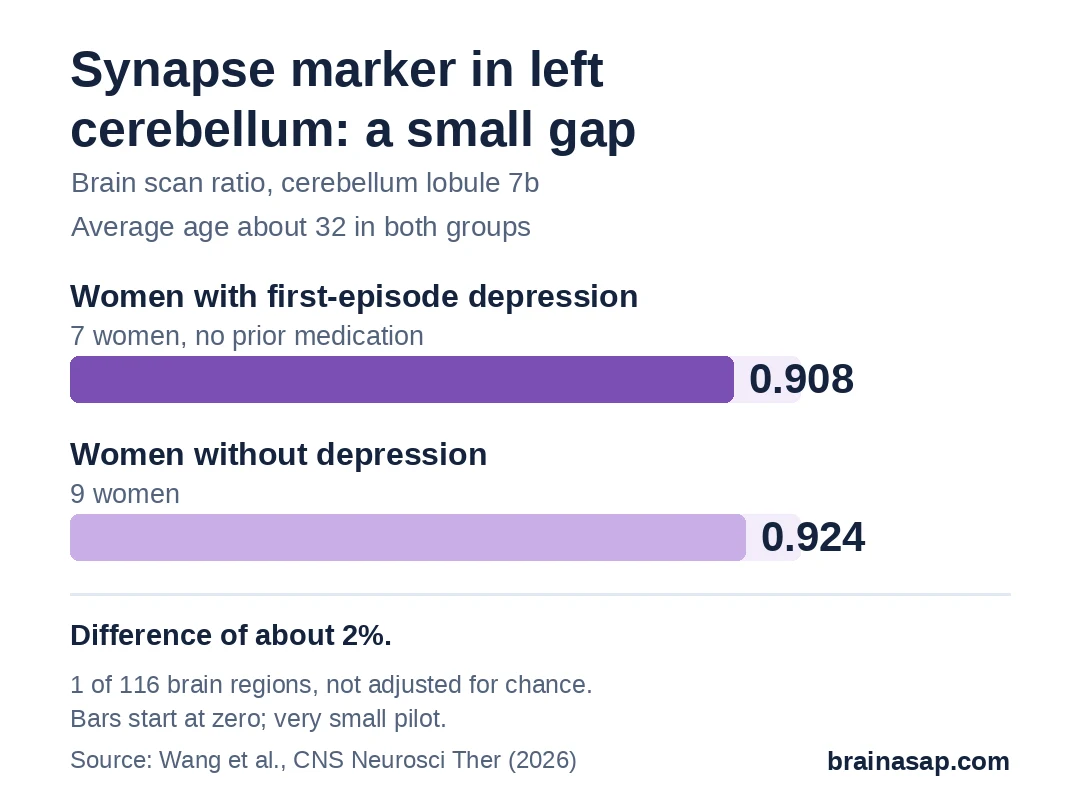
- 1 cerebellum spot was lower: synapse marker ratio of 0.908 in women with depression vs. 0.924 in women without, a gap of about 2%.
- Lower marker went with worse symptoms: across all 16 women, lower readings in that spot went with higher depression and anxiety scores.
- That spot looked less in step with the right frontal lobe: readings in the 2 areas matched up less closely in women with depression.
- Stressed mice showed lower readings too: brain-wide, with 9 stressed female mice and 9 unstressed ones.
Source: CNS Neuroscience & Therapeutics (2026) | Wang et al.
Depression is usually pictured as a problem in the front of the brain, where mood and decisions are handled. Far less attention goes to the cerebellum, a structure at the back best known for balance and coordination.
A small team of researchers in Hangzhou, China, wanted to look at it anyway. They asked whether the cerebellum shows a sign of depression early, before any medication has had a chance to change the picture.
How Do You Scan a Synapse?
Synapses are the tiny gaps where one brain cell passes a signal to the next. Researchers can't count them directly in a living person, but they can estimate how many there are using a PET scan.
Each woman got an injection of a radioactive tracer called [18F]UCB-H, which sticks to a protein found in synapses. A scanner then recorded how much tracer each part of the brain picked up. A higher reading in a region means more of the protein, and likely more synapses, there.
Scientists compare each region to a reference reading from the same scan, which gives a ratio. Ratios close to 1 are common, so differences can look tiny.
7 Women With Depression, 9 Without
All 16 participants were adult women. Those 7 women were having their first episode, scored 18 or higher on a standard depression interview (average about 23), and had never taken antidepressants. Women without depression, similar in age and education, averaged about 3.
Only women were included on purpose, since depression is about twice as common in women and previous research suggests it can look different in them. That also means nothing here says what happens in men.
One Cerebellum Spot Read Slightly Lower
Readings were compared across 116 brain regions. Only one stood out in the usual way: a region in the left cerebellum called lobule 7b.
- Women with depression: 0.908
- Women without depression: 0.924
That is a difference of roughly 2%. It was consistent enough within each group to look large statistically, but with only 16 women and 116 regions tested, a few false alarms are expected.
Women with lower readings in that spot also scored worse on the depression interview (correlation of −0.57) and an anxiety interview (−0.55). With so few women, correlations like these can swing a lot.
Did the Cerebellum Stop Talking to the Front of the Brain?
Next came a check of whether readings in cerebellum regions rose and fell together with readings in the frontal lobes. In women with depression, left cerebellum regions, including lobule 7b, matched up less with the right frontal lobe. That is called uncoupling.
A separate measure of how much brain regions work in step also dropped in the right frontal area. Connections within the cerebellum itself looked simpler in the women with depression.
A caution on the word talking: these tracer readings show which regions look alike, not that signals really travel less between them.
Mice Under Stress Showed Lower Readings Too
In a side experiment, 9 female mice were stressed by restraining them 4 hours a day for 21 days, then compared with 9 unstressed mice. Stressed mice spent more time motionless in a swim test and less time in the center of an open arena, and their brain scans showed lower tracer uptake.
This fits a link between stress and fewer synapses, but mouse stress is not human depression, and the mouse results were brain-wide rather than specific to the cerebellum.
Why 16 Women Can Only Suggest a Direction
- Very small groups: 7 and 9 women, from one hospital.
- Many comparisons: The cerebellum result was one of 116 regions, with no adjustment for chance findings.
- Snapshot only: A single scan can't show whether lower readings come before depression or follow it.
- Not a diagnostic test: Nothing here shows a scan can identify depression in one person.
- Women only: Findings may not apply to men.
A Lead for Larger Scans to Confirm
This pilot nominates the left cerebellum as a place worth checking. It doesn't show that depression thins out synapses there, or that treatments should target it.
Bigger studies need to answer whether the same small gap, and the weaker match with the right frontal lobe, shows up in a larger group of newly diagnosed women, and whether it changes with treatment.
Citation: DOI: 10.1002/cns.71144. Wang J, Lin Y, Zhang T, et al. Left cerebellar-right frontal uncoupling in female first-episode major depressive disorder: synaptic evidence from [18F]UCB-H PET. CNS Neurosci Ther. 2026;32:e71144.
Study Design: Exploratory cross-sectional PET imaging study with a small mouse side experiment.
Sample Size: 7 women with first-episode depression, 9 healthy women; 9 stressed and 9 control female mice.
Key Statistic: Left cerebellar lobule 7b tracer ratio 0.908 vs. 0.924 (p = 0.012, uncorrected for 116 regions).
Caveat: Very small, women-only pilot; hypothesis-generating; cross-sectional; mouse results do not establish human causation.