TL;DR: A person with severe inherited retina disease saw a wide blue glow each time a weak, humming magnetic field was aimed near the eyes, but nothing when it was aimed at the back of the head or switched off. It's a single-person experiment that points to surviving retina cells, not the brain, as the source.
Key Findings
- One person with advanced Stargardt disease and very weak retina responses.
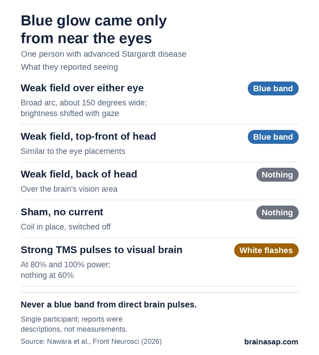
- Blue band seen with the coil over either eye or the forehead.
- Nothing seen with the coil over the back of the head or switched off.
- Direct brain zaps looked different: White flashes, not a blue band.
- Descriptions only: No measured brightness or color.
Source: Frontiers in Neuroscience (2026) | Nawara et al.
Put a slowly pulsing magnetic field near your head in a dark room, and you may see a flicker of light that isn't there. These "magnetophosphenes" are well documented, and in healthy people they're thought to start in the retina.
But what if the retina's light-sensing cells are mostly gone? Researchers at Western University in Canada tested one person with severe retina disease to find out.
A Retina With Few Working Light Sensors
The volunteer has advanced Stargardt disease, an inherited condition that destroys the light-sensing cells in the center of the retina. Electrical tests of the eye showed responses from those cells were severely weakened.
That made this person a useful test case. If a magnetic field still produced light, it would have to be acting on whatever retina wiring was left, or on the brain itself.
A Weak Field Aimed at the Eyes, Then the Head
The team used a coil that produces a magnetic field humming at 20 times a second, the rate where people see these flashes most easily. Each exposure lasted 5 seconds, in complete darkness. They moved the coil to four spots:
- Over the right eye
- Over the left eye
- Top-front of the head
- Back of the head: Over the brain's vision area.
Fake "sham" trials with the coil in place but no current were mixed in at random.
A Blue Band, but Only Near the Eyes
With the coil over either eye or the top-front of the head, the volunteer saw a large glowing band, curving across roughly 150 degrees of view. Every time, they called it blue. Looking up or down changed its brightness a little.
With the coil over the back of the head, and in every sham trial, they saw nothing.
Zapping the Brain Directly Looked Different
In a second session, the team used transcranial magnetic stimulation, or TMS, to send much stronger pulses into the brain's vision area. The electric field TMS creates in the brain is about 100 to 1,000 times stronger.
At the higher settings, the volunteer saw brief "white points" or "flashes," mostly on one side. They never saw a blue band. The researchers argue the blue glow must start in surviving cells deeper in the retina, which the brain then fills in.
One Person, Described in Words
- Single participant: No statistics, and it may not apply to others.
- Self-reported: No measured brightness, color matching or thresholds.
- Fixed order: Coil positions were always tested in the same sequence.
- Exact pathway unknown: Which retina cells respond can't be pinned down.
- One strength tested: No dose-response mapping.
Could the Leftover Retina Be Put to Use?
This isn't a treatment, and nobody's sight improved. What it suggests is that a badly damaged retina can still turn a very weak electrical nudge into conscious light. The next question is whether that holds in more people with retina disease, and whether it could someday help test how much of the retina still works.
Citation: DOI: 10.3389/fnins.2026.1893022. Nawara O, Fresnel E, Juncal V, Prato FS, Hutnik C, Bouisset N. Blue magnetophosphene perception under subthreshold magnetic stimulation in a human retinal degeneration model. Front Neurosci. 2026;20:1893022.
Study Design: Single-subject experimental study with sham, coil-position and TMS comparisons.
Sample Size: 1 participant with advanced Stargardt disease.
Key Statistic: 20 Hz, 100 mT RMS field evoked a blue arcuate band (~150°) with periorbital and frontal coil placement; none with occipital placement or sham; TMS at 80 to 100% output evoked achromatic phosphenes.
Caveat: Single participant; qualitative self-reports; no psychophysical measures.