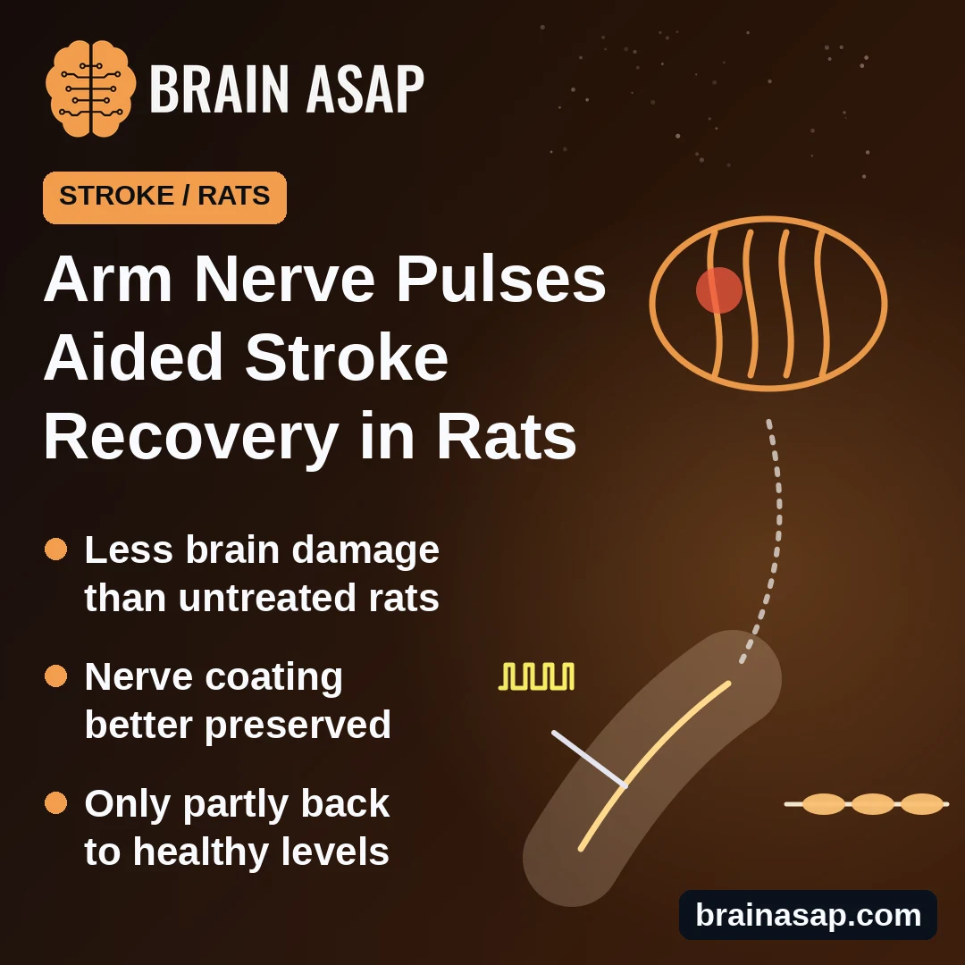
TL;DR: In a 2026 study, rats that had a stroke got 7 sessions of mild electrical pulses to the median nerve, which in people runs through the wrist. Compared with untreated stroke rats, they did better on movement tests, had less brain damage and kept more of the coating that insulates brain nerve fibers, though they did not fully recover.
Key Findings
- Better movement scores: stroke rats given nerve pulses had lower nerve-damage scores, stayed longer on a spinning rod and slipped less on a grid walk than stroke rats without pulses.
- Less brain damage: the area of dead brain tissue was smaller in stroke rats given pulses than in stroke rats without them.
- More nerve coating kept: myelin, the fatty coating that insulates nerve fibers, was better preserved in stroke rats given pulses.
- Only part of the way back: stroke rats given pulses still did worse on movement tests than rats that never had a stroke.
- A tiny RNA may be involved: blocking a small molecule called miR-1-3p partly copied the benefits; boosting it weakened them.
Source: Frontiers in Behavioral Neuroscience (2026) | Li et al.
Most people who have a stroke have trouble using one arm, and many still do months later.
One approach being tried is electrical stimulation of the median nerve, which runs down the forearm through the wrist. This nerve sends a strong stream of signals to the brain’s movement areas, so stimulating it might help the injured brain rewire.
A 2026 study in Frontiers in Behavioral Neuroscience, by researchers at the Second People’s Hospital of Kunming in China and Universiti Malaysia Sabah, tested this in rats and looked for how it might work.
How the Rats Were Treated
Researchers caused a stroke in adult male rats by blocking a major brain artery for 60 minutes, then reopening it. That mimics the most common type of human stroke, caused by a clot.
Starting 3 days later, some rats got median nerve stimulation in a front leg:
- How: a fine needle electrode placed at the nerve
- Dose: 1 milliamp, 2 pulses per second, for 20 minutes
- Schedule: every other day for 14 days, 7 sessions in all
The key comparison was between 3 groups of 18 rats each:
- Healthy rats: surgery without a stroke
- Untreated stroke rats: stroke, no stimulation
- Stimulated stroke rats: stroke plus nerve stimulation
Within each group, 6 rats went to each type of lab test, so most comparisons rest on 6 rats per group. Testers did not know which group each rat was in.
Stimulated Rats Moved Better and Had Less Brain Damage
After the 14 days of treatment, stimulated stroke rats did better than untreated stroke rats on 3 tests:
- Nerve-damage score: a 0-to-7 rating of problems such as a bent front leg when the rat is lifted by the tail. Stimulated rats scored lower, meaning fewer problems.
- Spinning rod: stimulated rats stayed on longer before falling.
- Grid walk: stimulated rats scored better on a test of foot slips.
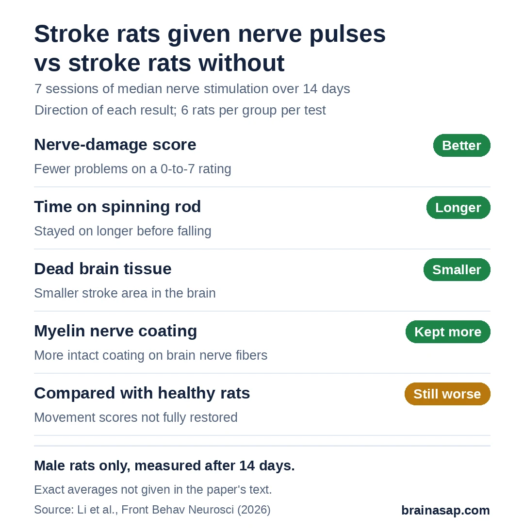
Stained brain slices showed a smaller area of dead brain tissue in stimulated rats, more surviving nerve cells, and fewer overactive immune cells.
But stimulation did not bring rats all the way back. Stimulated stroke rats still scored worse on movement tests than healthy rats. A check of the stimulated nerve itself found no signs of damage.
Nerve-Fiber Coating Held Up Better
Nerve fibers in the brain are wrapped in myelin, a fatty coating that lets signals travel quickly. Stroke strips some of it away.
Under a microscope, stimulated stroke rats had more intact myelin than untreated stroke rats, and more of a key myelin protein.
They also had more BDNF, a growth factor that helps nerve cells survive and helps build myelin, along with more of the receptor BDNF acts on.
A Tiny RNA Molecule May Link Stimulation to Repair
Researchers suspected a small RNA molecule called miR-1-3p, which can turn down BDNF production. In stimulated stroke rats, miR-1-3p levels were lower.
To test the link, other groups of rats got a brain injection 1 hour before the stroke:
- A miR-1-3p blocker: partly copied the benefits of stimulation, even without stimulation.
- A miR-1-3p booster: weakened several of the benefits of stimulation and lowered BDNF-pathway proteins.
That fits the idea that stimulation works partly by lowering miR-1-3p, which frees up BDNF. Li and colleagues described it as involvement “at least in part” and said direct targeting still needs confirming.
Limits of This Rat Study
- Rats, not people: rat strokes are induced and uniform, unlike strokes in people.
- Male rats only: results may differ in females.
- Short follow-up: outcomes were measured after 14 days, so lasting benefit is unknown.
- Small subgroups: most lab measurements used 6 rats per group.
- Injections before the stroke: the RNA blocker and booster were given before the stroke happened, which can’t happen in real patients.
Would Nerve Stimulation Help Repair the Brain in Stroke Patients?
This research group has already tried ultrasound-guided median nerve stimulation in a clinical study, which found better arm function soon after treatment.
What remains open is whether that benefit in people comes with the same myelin repair seen in these rats, and whether it lasts beyond the first weeks.
Citation: DOI: 10.3389/fnbeh.2026.1820913. Li R, Lu J, Zhang C, Zhang X, Wang M, Zhang P, Yang K, Fang H, Gao Y, Mohamed K, Che Noh CI, Li Y, Chin KL. Median nerve electrical stimulation promotes myelin regeneration through regulating miR-1-3p/BDNF/TrkB-MAPK pathway in a transient cerebral ischemia rat model. Front Behav Neurosci. 2026;20:1820913.
Study Design: Randomized, blinded-assessment rat experiment: transient middle cerebral artery occlusion (60 min) with median nerve electrical stimulation (7 sessions over 14 days) and intracerebroventricular miR-1-3p antagomir/agomir.
Sample Size: 144 adult male Sprague-Dawley rats in 8 groups of 18 (6 per analysis type).
Key Statistic: Stimulation vs untreated stroke: lower neurological severity score and longer rotarod time (p < 0.01); smaller infarct (p < 0.001).
Caveat: Male rats only; 14-day follow-up; exact group means not reported in text; miR-1-3p targeting not directly confirmed.