TL;DR: In a Finnish brain-scan study of 40 adults with attention-deficit/hyperactivity disorder and 36 adults without it, activity in the front of the brain and a deep brain center was less closely linked in adults with ADHD during memory games. Rest and film scans showed no clear difference in those links.
Key Findings
- Weaker teamwork during memory games: While remembering numbers or square positions from a few steps back, adults with ADHD showed less coordinated activity between the frontal lobes and the striatum, a deep center for movement, habits and reward.
- No clear difference at rest or during a film: The scans did not show a clear difference in these brain links at rest or during a film with distracting sounds.
- Wiring result was shaky: One nerve-fiber pathway from the front of the brain looked weaker in ADHD, but the result disappeared when the team changed how the pathways were traced.
- Similar memory-game accuracy: Both groups got about the same share of answers correct, and adults with ADHD often answered faster.
Source: Human Brain Mapping (2026) | Tolonen, Roine and colleagues
For decades, ADHD research has pointed to a set of loops connecting the front of the brain with the striatum, a deep brain structure involved in movement, habits and reward. But these loops come in many separate strands, and people's brains differ.
Which strands are affected in adults with ADHD, and when? A 2026 study in Human Brain Mapping, by a team from Aalto University and the University of Helsinki, mapped those loops person by person to find out.
Mapping Each Person's Brain Loops
Instead of using a standard brain map for everyone, the team traced each participant's own nerve-fiber pathways from 6 parts of the striatum on each side to the front of the brain. That gave a personal set of loops for every person.
Researchers compared 2 groups of Finnish adults:
- Adults with ADHD (40 people): diagnosed by psychiatrists, without other major psychiatric or neurological conditions
- Adults without ADHD (36 people): similar in age, gender, education and general thinking ability
Of the adults with ADHD, 33 took stimulant medication and stopped it for 24 hours before the scan.
What Did People Do in the Scanner?
The study compared 4 activities in the scanner:
- A number memory game: numbers flashed one at a time, and people pressed a button to say whether each matched the one shown 1, 2 or 3 steps earlier
- A location memory game: the same idea, using the position of a white square in a grid
- Rest: looking at a cross on a screen for about 7 minutes
- A film with distractions: watching a 14-minute clip from a Finnish film while ignoring loud background sounds, followed by a quiz
A wiring scan also measured the nerve-fiber pathways themselves.
Weaker Teamwork During the Memory Games
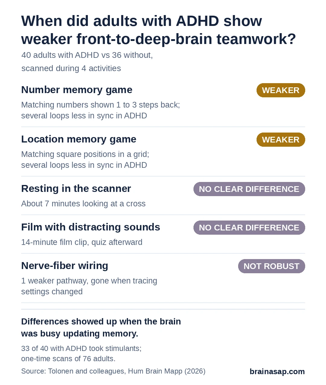
During both memory games, adults with ADHD showed less coordinated activity between the striatum and several frontal areas. These included regions for planning and holding information in mind, for movement, and for weighing rewards.
At rest and during the film, the 2 groups showed no clear difference in these loops.
Tolonen and colleagues suggested the differences appear when the striatum is actively needed. Memory games that constantly swap old information for new seem to call on these loops in a way that rest and film-watching do not.
Did the Memory Games Go Worse?
Not by accuracy. Both groups made more mistakes as the games got harder, but they hit about the same share of correct matches at every level. Adults with ADHD often responded faster, especially in the location game.
On a separate memory test outside the scanner, where people recalled the last few positions in a long sequence, adults with ADHD did score lower.
Limits of a 76-Person Study
- Medication mixed in: 33 of 40 adults with ADHD used stimulants, so effects of ADHD and of long-term medication could not be fully separated, even with a 24-hour break
- Fragile wiring finding: the one weaker nerve-fiber pathway did not hold up under different analysis settings
- A narrow group: people with depression, anxiety, learning disorders or other conditions were excluded, though these often come with ADHD
- Modest size: smaller effects may have been missed after correcting for many comparisons
What This Means for Understanding Adult ADHD
The study suggests that in adults, the well-known front-to-striatum loops may not look different all the time. They looked different when the brain was busy updating information in memory.
A useful next step would be to test the same personal loop maps in adults with ADHD who have never taken stimulants, and in larger groups that include common conditions like anxiety and depression.
Citation: DOI: 10.1002/hbm.70652. Tolonen T, Laine M, Alho K, Leppämäki S, Tani P, Koski A, Salmi J, Roine T. Individualised structural and functional frontostriatal connectivity mapping in adult ADHD. Human Brain Mapping. 2026;47:e70652. PMID: 42823824.
Study Design: Cross-sectional case-control multimodal 3 T MRI study with individually tractography-defined frontostriatal circuits (6 striatal seeds per hemisphere); diffusion MRI structural connectivity and microstructure; functional connectivity during digit and visuospatial n-back, resting state and a naturalistic film task with distractors; FDR correction.
Sample Size: 40 adults with ADHD and 36 neurotypical controls in the memory and film analyses. Resting-state analysis included 35 and 34, respectively.
Key Statistic: Reduced frontostriatal functional connectivity in ADHD during digit n-back (for example left dorsal putamen circuit t(74) = −3.08, p = 0.001) and visuospatial n-back (left dorsal putamen t(74) = −3.24, p < 0.001), FDR-corrected; no clear group differences in these links at rest or during the film.
Caveat: Most ADHD participants used stimulants; structural result not robust to threshold choice; comorbidities excluded; modest sample.