TL;DR: A 2026 review summed up years of studies that filmed brain cells lighting up in living mice bred to develop Alzheimer’s-like disease. Some cells fired far more than normal, especially near the protein clumps typical of Alzheimer’s, while others went silent. The review argued that AI tools could pick up network-wide changes that simple activity counts miss.
Key Findings
- Overactive and silent cells side by side: in one widely cited study of Alzheimer’s-model mice, about 21% of brain cells in the outer brain were overactive and about 29% were silent.
- Overactive cells clustered near plaques: cells firing too much were found mostly close to amyloid plaques, the protein clumps that build up in Alzheimer’s.
- Tau pushed the other way: in mice with tau buildup, the second Alzheimer’s protein, many cells went quiet, even when amyloid was also present.
- Memory-map cells became unreliable: in some Alzheimer’s-model mice, cells that normally fire in one spot of an enclosure fired less consistently or all over the place.
- AI as a sharper readout: the review argued that AI methods, including the authors’ own, could detect disease patterns in whole networks of cells and later test whether treatments restore them. That use is still mostly untested.
Source: Frontiers in Aging Neuroscience (2026) | Gerasimov et al.
Long before memory fails in Alzheimer’s disease, brain cells start talking to each other the wrong way. Some get too loud, others fall quiet.
Scientists can watch this happen in mice. A 2026 review in Frontiers in Aging Neuroscience, by researchers at Peter the Great St. Petersburg Polytechnic University and the Pavlov Institute of Physiology in Russia, pulled together what those recordings show and how best to analyze them.
How Researchers Watch Brain Cells Fire
When a nerve cell fires, calcium rushes inside it. Mice can be given a gene for a protein that glows when calcium rises, so active cells light up under a microscope.
There are 2 main setups:
- A large microscope: the mouse’s head is held still, which gives sharp images of single cells but limits what the mouse can do.
- A miniscope: a tiny microscope worn on the mouse’s head, so it can explore freely while cells deep in the brain are recorded, though images are blurrier.
Mice in these studies are bred to carry human gene changes that cause amyloid plaques, tau tangles or both, the 2 protein buildups that define Alzheimer’s in people.
Some Brain Cells Fire Too Much, Others Go Silent
The most consistent finding across studies was a mix of extremes. In one widely cited study of mice with amyloid plaques, aged 6 to 10 months:
- About 21% of cells in the outer layer of the brain fired far more than normal.
- About 29% were silent.
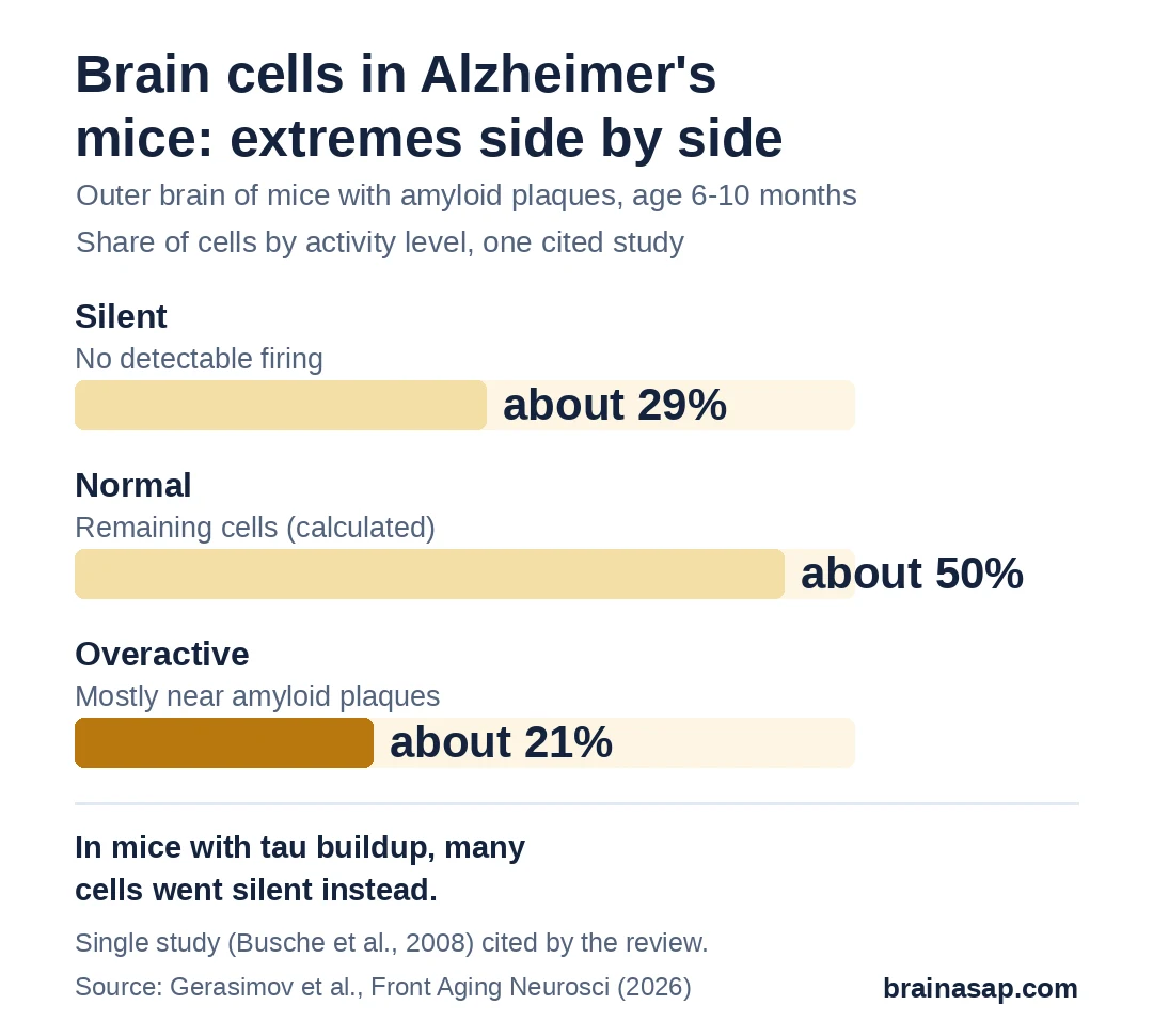
Overactive cells sat mostly near amyloid plaques. Later studies that filmed the same cells over weeks found that overactivity built up gradually: in one, about 21% of cells that started with middling activity became overactive over time.
Plaques weren’t required, though. Mice carrying only a presenilin gene change, which never form plaques, also had overactive cells. Restoring how cells store calcium brought their activity back toward normal.
Tau Protein Had the Opposite Effect
Tau, the protein that forms tangles inside cells in Alzheimer’s, pushed brain activity the other way. In mice with tau buildup, many cells showed no detectable firing at all.
When mice had both amyloid and tau, the quieting effect of tau won out, and activity across the outer brain dropped. This came from loose tau, not the larger tangles.
That matters for anyone measuring brain activity: the same disease can look like too much activity or too little, depending on which protein dominates at that stage.
Memory Circuits Lost Their Precision
Miniscope studies in freely moving mice looked at the hippocampus, a memory hub. It holds “place cells” that normally fire when a mouse is in one particular spot.
- In one model: place cells were less active and fired less consistently in the same spot from one trip to the next.
- In another model: cells fired more often, but spread their firing across the enclosure instead of in one spot.
Either way, the mental map got blurrier. Other studies found that a type of cell that normally calms the network was overactive while another calming type was underactive, throwing off the balance between excitation and calm.
Why the Review Pushes for AI in the Analysis
Most studies boil recordings down to simple counts: how often each cell fires, and how often pairs of cells fire together. Gerasimov and colleagues argued that such counts miss how hundreds of cells work together as a network.
They described newer approaches, including methods that compress a whole network’s activity into a simple shape, and AI models similar to those behind chatbots, which learn patterns across many cells and moments at once.
Their own AI model, trained on a large public set of mouse recordings, could tell recordings of Alzheimer’s-model mice from normal mice.
In another of their studies, a compound they are developing appeared to shift network patterns in Alzheimer’s-model mice toward those of normal mice, though they noted that such shapes are not direct proof of recovery.
What the Review Doesn’t Settle
- Mice, not people: each mouse model copies only part of Alzheimer’s, and results differed by model, age and brain area.
- Not a systematic review: studies were hand-picked rather than found through a set search, and results were not combined statistically.
- Authors reviewing their own tools: the AI model and the test compound described as promising come from the review team’s own labs.
- AI results can shift with settings: the same recording can produce different network shapes depending on how the analysis is set up, a limitation the review acknowledged.
Could Brain-Network Patterns Become a Test for Alzheimer’s Treatments?
That is the review’s main proposal: use network patterns from these recordings to judge whether a treatment restores normal brain activity in mice, and at what stage it’s too late.
Testing that idea will take shared data sets, agreed-upon methods, and experiments that change cell activity directly to confirm what the AI models flag.
Citation: DOI: 10.3389/fnagi.2026.1930851. Gerasimov E, Fedorov E, Bezprozvanny I, Pchitskaya E. Dissecting neuronal circuit function and dysfunction in Alzheimer’s disease mouse models: from conventional calcium imaging analysis to machine learning-based approaches. Front Aging Neurosci. 2026;18:1930851.
Study Design: Narrative review of in vivo calcium imaging (two-photon and miniscope) studies in Alzheimer’s disease mouse models and of analysis methods, including manifold learning and transformer-based AI.
Sample Size: Not applicable (narrative review; mouse models include APP/PS1, PS45, 5xFAD, 3xTg-AD, rTg4510).
Key Statistic: In APP/PS1 mice aged 6–10 months, about 29% of cortical neurons were silent and about 21% hyperactive, with hyperactive cells near plaques (Busche et al., 2008, as summarized).
Caveat: Mouse models only; selective narrative review; authors discuss their own AI model and compound.