TL;DR: German researchers gave 16 rats 45 minutes of electrical stimulation through a wire placed in the front of the brain. In rats whose mothers had an immune reaction during pregnancy, a common animal model of schizophrenia, a deeper reward area responded less in the first minute afterward than in ordinary rats. This short-lived difference comes from a small early experiment.
Key Findings
- Different front-brain rhythms: across the recording, immune-model rats, whose mothers got an immune-triggering injection in pregnancy, had a much smaller share of 2 brainwave types in the front of the brain than control rats.
- Firing increased in both groups: on average, recorded nerve cells fired more in the first minute after stimulation in all four sampled brain areas.
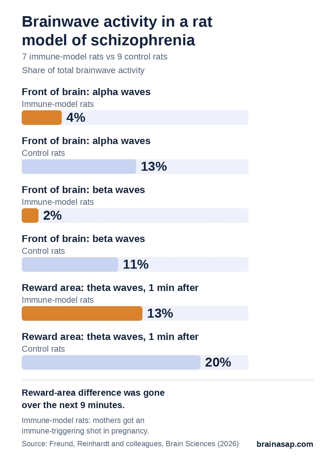
- Weaker reward-area rhythm: in that first minute, slow theta waves made up 13% of brainwave activity in a reward area of immune-model rats, compared with 20% in control rats.
- Gone within minutes: researchers detected no clear difference on that measure over the next nine minutes.
Source: Brain Sciences (2026) | Freund, Reinhardt, Juckel and colleagues
Up to 1 in 3 people with schizophrenia don't get better on standard antipsychotic medicines. Deep brain stimulation, which sends steady electrical pulses through a thin wire implanted in the brain, is one idea being explored for them.
So far it has been tried in only a handful of patients, so researchers are turning to animals to learn what the stimulation actually does to brain activity.
How the Rats Were Tested
A 2026 study in Brain Sciences, from a team at Ruhr University Bochum in Germany, compared 2 groups of adult male rats:
- Immune-model rats (7): their mothers got an injection during pregnancy that mimics a viral infection. Infection in pregnancy is a known risk factor for schizophrenia, and the grown offspring show schizophrenia-like behavior and brain changes.
- Control rats (9): their mothers got a salt-water injection instead.
Under anesthesia, each rat had a stimulating wire placed in the medial prefrontal cortex, a front-of-brain area involved in planning and self-control. It received 45 minutes of rapid pulses, the same pattern used in earlier rat studies that improved schizophrenia-like behavior.
Recording wires picked up brainwaves and the firing of single nerve cells in 4 areas, including the prefrontal cortex and the nucleus accumbens, a deep area involved in reward and motivation. Recordings ran for 10 minutes before stimulation and 10 minutes after.
How Did Immune-Model Rats Differ Overall?
Brainwaves come in speed bands, from slow theta waves to faster alpha, beta and gamma waves. Researchers measured what share of total brainwave activity each band made up, before and after stimulation.
In the usable prefrontal recordings, immune-model rats had a smaller overall share of alpha waves (4% vs 13%) and beta waves (2% vs 11%) than control rats. Lower alpha and beta activity has also been reported in people with schizophrenia.
Nerve cells in the hippocampus, a memory area, also fired less often overall in immune-model rats: about 270 vs 720 signals per minute.
What Changed After Stimulation?
Right after the 45 minutes of stimulation, nerve cells in all 4 areas fired more and in more bursts, in both groups. Over the following 9 minutes, firing no longer differed from where it started.
2 differences between the groups showed up only in that first minute:
- Nucleus accumbens: theta waves made up 13% of brainwave activity in immune-model rats vs 20% in control rats, though the groups had not differed before stimulation
- Hippocampus: nerve cells fired about 710 vs 1,870 times per minute
The team read this as a weaker response to prefrontal stimulation in 2 deeper brain areas of immune-model rats. Weak theta rhythms have been linked to problems with mental control in people with schizophrenia, so a blunted response could matter if it holds up.
Why These Results Are Early
- 16 rats: the team called the work exploratory and said its statistics were prone to false positives
- No sham group: without rats that got wires but no stimulation, changes after stimulation could partly come from more time under anesthesia
- Anesthetized rats: one of the anesthetics, ketamine, is itself used to mimic schizophrenia in animals, and both drugs alter brainwaves
- Males only, left side only: results may differ in females or in the other half of the brain
- No behavior measured: the study recorded brain signals, not whether the rats behaved differently
A Possible Brain Signal to Track
If larger studies confirm it, theta waves in the nucleus accumbens could become a measurable sign of whether prefrontal stimulation is reaching the brain circuits involved in schizophrenia.
Whether deep brain stimulation can help people whose schizophrenia resists medicine is still an open question that only careful human trials can answer.
Citation: DOI: 10.3390/brainsci16101003. Freund N, Reinhardt PR, Abraham M, Funke K, Juckel G. Deep Brain Stimulation Modulates Nucleus Accumbens Theta-Band Power in a Rodent Model of Schizophrenia. Brain Sciences. 2026;16(10):1003. PMID: 42939257.
Study Design: Exploratory animal study; maternal immune activation (PolyI:C, gestational day 15) rat model; 45 min of 130 Hz prelimbic mPFC deep brain stimulation under urethane/ketamine anesthesia; local field potentials and single-unit recordings in mPFC, nucleus accumbens, ventral hippocampus and ventral tegmental area for 10 min before and after.
Sample Size: 16 adult male Wistar rats (7 MIA offspring, 9 controls); LFP data lost for 3 rats.
Key Statistic: NAc relative theta power 60 s after DBS: MIA 0.133 vs control 0.197 (group x time interaction F(2,20) = 4.96, p = 0.018; post hoc p = 0.005). mPFC relative alpha 0.044 vs 0.126 (p = 0.009) and beta 0.018 vs 0.106 (p < 0.001).
Caveat: Very small exploratory sample; no sham-stimulation group; anesthetized animals (ketamine is an NMDA antagonist); males and left hemisphere only; no behavioral outcomes.