TL;DR: In 32 healthy young adults, fast brain waves recorded during 2.5 minutes of rest were linked to how well people later remembered names paired with faces. More fast activity over the front of the head and less over the back went with better recall.
Key Findings
- Fast rest waves linked to recall: High-gamma activity explained about 38% of differences in delayed name recall.
- Front and sides positive, back negative: Opposite links depending on where on the head.
- Only 1 of 9 models held up: Others faded after correcting for multiple tests.
- Slower theta waves showed no clear link.
- Brain-network syncing: Hints only, none significant after correction.
Source: Cognitive Neurodynamics (2026) | Scianatico et al.
Forgetting someone's name right after being introduced is common, and it gets more common with age. Matching a face to a name pulls together vision, language and memory at once, which is why doctors use face-name tests to spot early memory decline.
Researchers at the University of Bari in Italy asked whether the brain's activity while doing nothing at all, before any test starts, says anything about how well a person will learn new face-name pairs.
A Short Rest Recording, Then a Name Test
Thirty-two healthy adults (average age 23; 19 women, 13 men) sat with eyes closed for 2.5 minutes while a 19-electrode EEG cap recorded their brain waves. Then they took the Italian Face-Name Association Test:
- Learning: 16 faces, each shown with a name to read aloud.
- Immediate recall: Name each face right away.
- Delayed recall: After 15 minutes of other tasks, name each face again. If they couldn't, they picked from 3 options.
The researchers focused on three rhythms: slow theta waves (4 to 7 cycles per second), low gamma (30 to 49) and high gamma (51 to 100), measured over the front, sides, center and back of the head.
High Gamma Stood Out for Delayed Recall
Of the nine combinations of rhythm and recall score, only one held up after correcting for running many tests: high-gamma activity and delayed total recall (free recall plus multiple-choice).
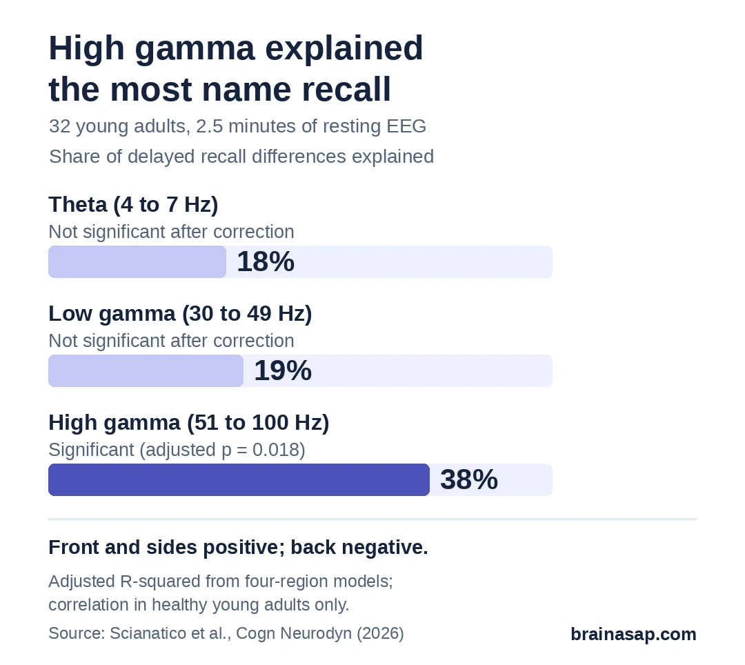
That model explained about 38% of the spread in scores between people. The direction depended on location:
- Front of the head: More high gamma, better recall.
- Sides (temporal areas): More high gamma, better recall.
- Back of the head: More high gamma, worse recall.
This front-positive, back-negative pattern matches an earlier study by the same group using general memory tests.
Checks Against Muscle Noise and Short Recordings
Very fast EEG signals can be contaminated by tiny muscle movements, especially near the temples and jaw. The researchers removed muscle-related signals and reran the model without the side regions. It still held, with the front positive and the back negative.
On average, only about 96 seconds of clean recording per person remained after cleanup. When the 4 people with less than a minute of clean data were dropped, the link got stronger, not weaker.
A Small Group of Young Adults and One Snapshot
- 32 people, all young and healthy: Results may differ in older adults or people with memory problems.
- Correlation only: Rest activity came first, but that does not show it causes better memory.
- Many top scores: 11 of 32 people got a perfect delayed total recall score.
- Short recording: 2.5 minutes, with some data lost to cleanup.
- Regions overlap: Brain signals from neighboring regions were fairly correlated with each other.
Could a Resting Brain Scan Flag Memory Changes?
A quick, eyes-closed EEG is cheap and easy, so a resting pattern that tracks memory could one day help screen for early memory decline. This study is a first step in healthy young people, not a test anyone can use.
The next question is whether the same front-versus-back gamma pattern shows up, and predicts memory, in larger groups of older adults and people with early memory problems.
Citation: DOI: 10.1007/s11571-026-10551-y. Scianatico G, Scaramuzzi GF, Çağlayan E, et al. Before the task begins: linking resting-state gamma oscillations to face-name associative memory. Cogn Neurodyn. 2026;20:169.
Study Design: Cross-sectional correlational study; eyes-closed resting EEG before the Italian Face-Name Association Test.
Sample Size: 32 healthy adults (mean age 23.3).
Key Statistic: High-gamma power in four regions predicted delayed total recall, adjusted R² = 0.378, FDR-adjusted p = 0.018 (frontal β = 0.99, temporal β = 0.64, posterior β = −1.16).
Caveat: Small young sample, short recording, ceiling effects; correlational only.