
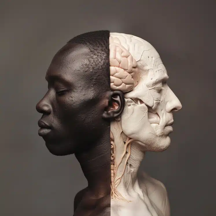
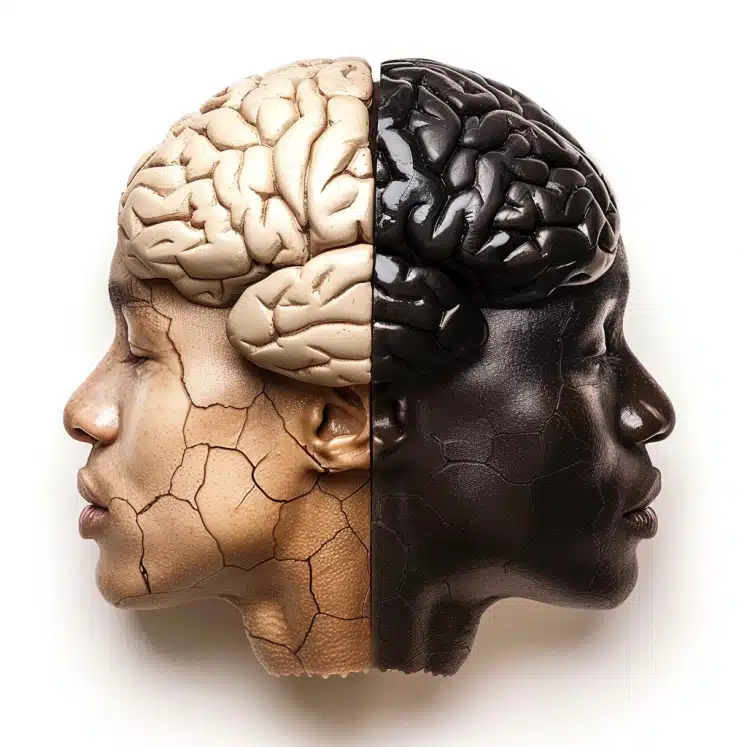
This study reveals significant differences in brain morphology between neurotypical young adults identified as White or African American, highlighting the impact of racial identity on brain structure.
Highlights:
- Brain Volume Differences: Significant volumetric differences were found in white matter, subcortical regions, and the choroid plexus between White and African American participants.
- Cortical Thickness Variability: Differences in cortical thickness were observed in various frontal, parietal, temporal, and occipital brain regions.
- Surface Area Disparities: Significant disparities in cortical surface area were identified between the racial groups across multiple brain regions.
Source: Frontiers in Integrative Neuroscience (2023)
Study Overview: Racial Identity (Black vs. White) in U.S. & Brain Morphology (2023)
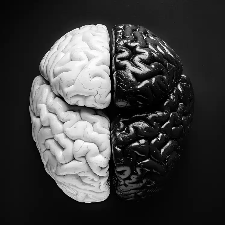
The study aimed to identify brain morphological variability due to racial identity in representative samples of neurotypical young adults.
The goal was to investigate differences in brain structure between White and African American participants.
Sample
- Participants: Data were analyzed from the Human Connectome Project (HCP) database.
- Racial Groups: Participants identified as White (n = 338) and African American (n = 56).
- Criteria: The sample included neurotypical individuals, excluding those with psychiatric symptoms, substance use disorders, endocrine disorders, irregular menstrual cycles, or neurological abnormalities.
Methods
- Data Source: HCP database provided demographic, clinical, psychiatric, and morphometric brain information.
- Morphometric Measures: Brain volumetry, cortical thickness, and cortical surface area were measured using MRI data.
- Statistical Analysis: Non-parametrical permutation analysis of covariance (ANCOVA) was conducted, adjusting for age, sex, education, and economic income. Results were adjusted for multiple comparisons using the family-wise error rate method.
- Tools: R version 3.6.3 and RStudio software version 1.2.5033 were used for data analysis.
Limitations
- Sample Imbalance: The study faced a significant imbalance in sample size between White (n = 338) and African American (n = 56) participants, limiting the statistical power.
- Exclusion Criteria: Implementing exclusion criteria for confounding variables maintained the imbalance and reduced sample size.
- Self-Identification: Racial identity was based on self-identification rather than genetic ancestry, which may affect the accuracy of the findings.
- Generalizability: Findings are specific to neurotypical young adults and may not generalize to other age groups or populations with cognitive impairments.
Most Likely Reasons for Brain Differences Between White & Black Americans
1. Genetic & Epigenetic Factors
Genetic Ancestry: Genetic differences may underlie many of the observed brain structural differences. Variations in genes related to brain development and function could be more prevalent in one racial group than another.
Epigenetics: Environmental factors can cause changes in gene expression without altering the DNA sequence. Stress, diet, and exposure to toxins can lead to epigenetic modifications that affect brain development and structure.
2. Socioeconomic & Educational Factors
Socioeconomic Status (SES): Lower SES is associated with reduced access to resources, which can impact brain development. African Americans in the study reported lower economic income and fewer years of education compared to Whites, reflecting broader societal inequalities.
Educational Opportunities: Differences in educational attainment can affect cognitive stimulation and brain development. Whites generally had more years of education, potentially leading to differences in brain structure due to varied cognitive demands and learning environments.
Potential Advantages & Disadvantages of the Respective Morphologies (Blacks & Whites)
1. White Participants
Advantages
- Larger Cortical Surface Area in Frontal & Parietal Regions: This may provide enhanced abilities in complex cognitive tasks, executive functions, and spatial reasoning, supporting better problem-solving skills and higher-level cognitive processing.
- Greater Cortical Thickness in Certain Areas: Thicker cortices in regions such as the superior temporal sulcus and rostral anterior cingulate cortex might be associated with better emotional regulation, social cognition, and decision-making.
Disadvantages
- Increased Brain Volume in Certain Regions: Larger volumes in some brain regions could potentially be linked to a higher susceptibility to certain neurological conditions, such as overactivity in areas related to anxiety and stress responses.
2. Black American Participants
Advantages
- Greater White Matter Volume: Increased white matter volume can enhance connectivity between different brain regions, potentially supporting more efficient communication within the brain and faster processing speeds.
- Larger Subcortical Structures: Larger volumes in subcortical regions like the caudate and thalamus may benefit motor control, coordination, sensory processing, and emotional regulation.
Disadvantages
- Smaller Cortical Surface Area in Some Regions: Reduced surface area in regions such as the lateral occipital cortex and inferior temporal gyrus could be linked to limitations in visual processing and certain aspects of memory and object recognition.
- Thinner Cortices in Various Brain Areas: Thinner cortices in areas like the cuneus and occipital cortex might be associated with reduced capabilities in processing visual information and potentially lower efficiency in certain cognitive tasks.
Conclusion: Whites vs. African Americans & Brain Structure
This study highlights significant differences in brain morphology between neurotypical young adults identified as White and African American, emphasizing the influence of racial identity on brain structure.
These findings underscore the critical impact of genetics, socioeconomic status, educational opportunities, environmental factors on brain development.
By identifying volumetric, cortical thickness, and surface area disparities, the research points to the necessity of including diverse racial groups in neuroimaging studies to ensure comprehensive and accurate understanding of human brain variability.
Addressing these differences is essential for developing personalized clinical treatments and reducing health disparities.
The study’s results call for increased representation of minority populations in biomedical research to avoid generalization bias and improve the effectiveness of medical interventions across all racial groups.
Overall, recognizing and accounting for brain morphological diversity can enhance the precision and inclusivity of neuroscience and healthcare practices.
References
- Study: Brain morphological variability between whites and African Americans: the importance of racial identity in brain imaging research (2023)
- Authors: Daniel Atilano-Barbosa & Fernando A Barrios


