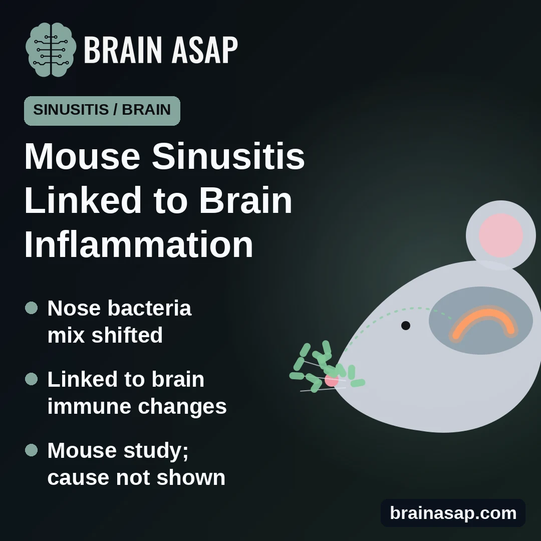
TL;DR: In mice with a long-lasting allergic-type sinus inflammation, the mix of nose bacteria changed, and signs of inflammation rose in the hippocampus, a memory area of the brain. Certain nose bacteria tracked with those brain changes, but the study cannot show they caused them.
Key Findings
- Nose and gut bacteria shifted: Overall community makeup differed from healthy mice, though variety stayed the same.
- Brain immune cells stirred: A microglia marker rose in both the smell center and the hippocampus.
- Inflammatory signal up in memory area: Il-6 gene activity rose in the hippocampus.
- Nose bacteria, not gut bacteria, tracked the brain: Several nasal families were linked to hippocampal gene activity.
Source: Frontiers in Immunology (2026) | Imai et al.
Chronic sinusitis is more than a stuffy nose. People who have it for months report more depression, anxiety and trouble thinking, and no one is quite sure why.
A team at Kyorin University in Tokyo wondered whether the bacteria living in an inflamed nose might be part of the story.
A Mouse Version of Stubborn Sinus Disease
The researchers modeled eosinophilic chronic rhinosinusitis, a hard-to-treat form driven by allergy-type inflammation and often linked to nasal polyps. Adult male mice were first primed with a skin treatment and the egg protein ovalbumin on one ear for 2 weeks, then had ovalbumin dripped into one nostril for 7 days. Control mice got inactive solutions.
The treated mice developed the expected nose changes: many eosinophils (an allergy-related white blood cell), more repair-type immune cells and extra mucus. Their weight, food and water intake stayed normal.
Nose and Gut Bacteria Both Changed
The researchers sequenced bacterial DNA from nasal tissue (7 control and 9 sinusitis mice) and from the gut (9 and 11). In both places, overall variety was unchanged, but the mix of bacteria differed between groups. Some families, including Staphylococcaceae, were more common in inflamed noses.
Signs of Inflammation in a Memory Area
In separate groups of 13 control and 14 sinusitis mice, the researchers looked at two brain regions: the olfactory bulb, which handles smell, and the hippocampus, which is central to memory.
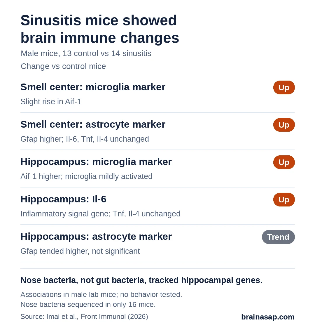
In the smell center, immune cells called microglia and support cells called astrocytes looked more active. In the hippocampus, microglia appeared mildly activated, and the inflammatory signal gene Il-6 rose.
Nose Bacteria Tracked the Brain Changes
When the researchers checked which bacteria moved together with brain gene activity, the nose stood out. Several nasal bacterial families, including Staphylococcaceae, Pseudomonadaceae and Neisseriaceae, rose and fell with hippocampal microglia, astrocyte or Il-6 markers. No gut bacteria showed such links in the hippocampus.
That points to the nose, which sits right next to the brain's smell pathways, as a possible route. But it is a link, not proof.
Why This Stops Short of Cause and Effect
- Associations only: No experiment changed the bacteria to test their effect.
- Male mice only: Sex and age differences were not studied.
- Clean lab mice: Their microbes differ from those in human noses.
- Small groups: Nose bacteria were sequenced in only 7 control and 9 sinusitis mice.
- No behavior tests: The study did not check whether memory or mood changed.
Could Treating the Nose Help the Brain?
The findings fit with reports that people with long-term sinusitis have more mood and thinking complaints. They do not show that sinus bacteria harm the brain.
The next question is whether changing the nasal bacteria, with antibiotics, probiotics or other treatments, actually calms inflammation in the hippocampus and protects memory and mood.
Citation: DOI: 10.3389/fimmu.2026.1811570. Imai R, Sugimoto Y, Osaki T, Hasegawa-Ishii S. Dysbiosis of nasal microbiota associated with hippocampal neuroimmune alterations in a mouse model of eosinophilic chronic rhinosinusitis. Front Immunol. 2026;17:1811570.
Study Design: Mouse model study with 16S rRNA sequencing of nasal and gut bacteria, brain gene expression and association analysis (MaAsLin2).
Sample Size: Male mice; microbiota 7 vs 9 (nose) and 9 vs 11 (gut); brain 13 vs 14.
Key Statistic: Nasal community makeup differed between groups (PERMANOVA R² = 0.183, p = 0.021).
Caveat: Associations only; male lab mice; no behavior measured.