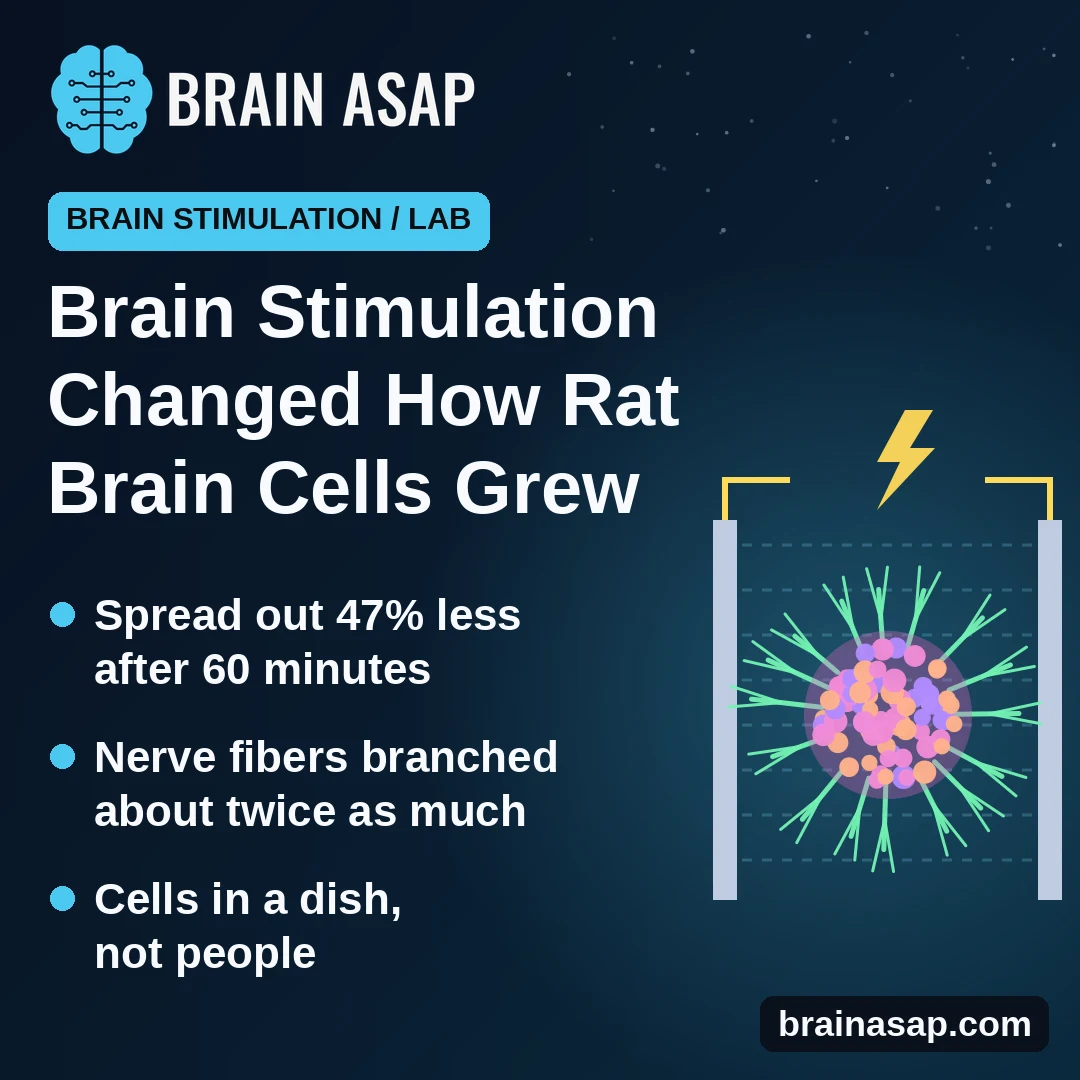
TL;DR: In a 2026 lab study, researchers ran a steady electric current for 15 or 60 minutes through tiny balls of rat brain cells grown in a dish. After 60 minutes, the balls spread outward about half as much over 3 days while their nerve fibers grew longer and more branched: an early clue to how brain stimulation reshapes tissue, not evidence about treatments in people.
Key Findings
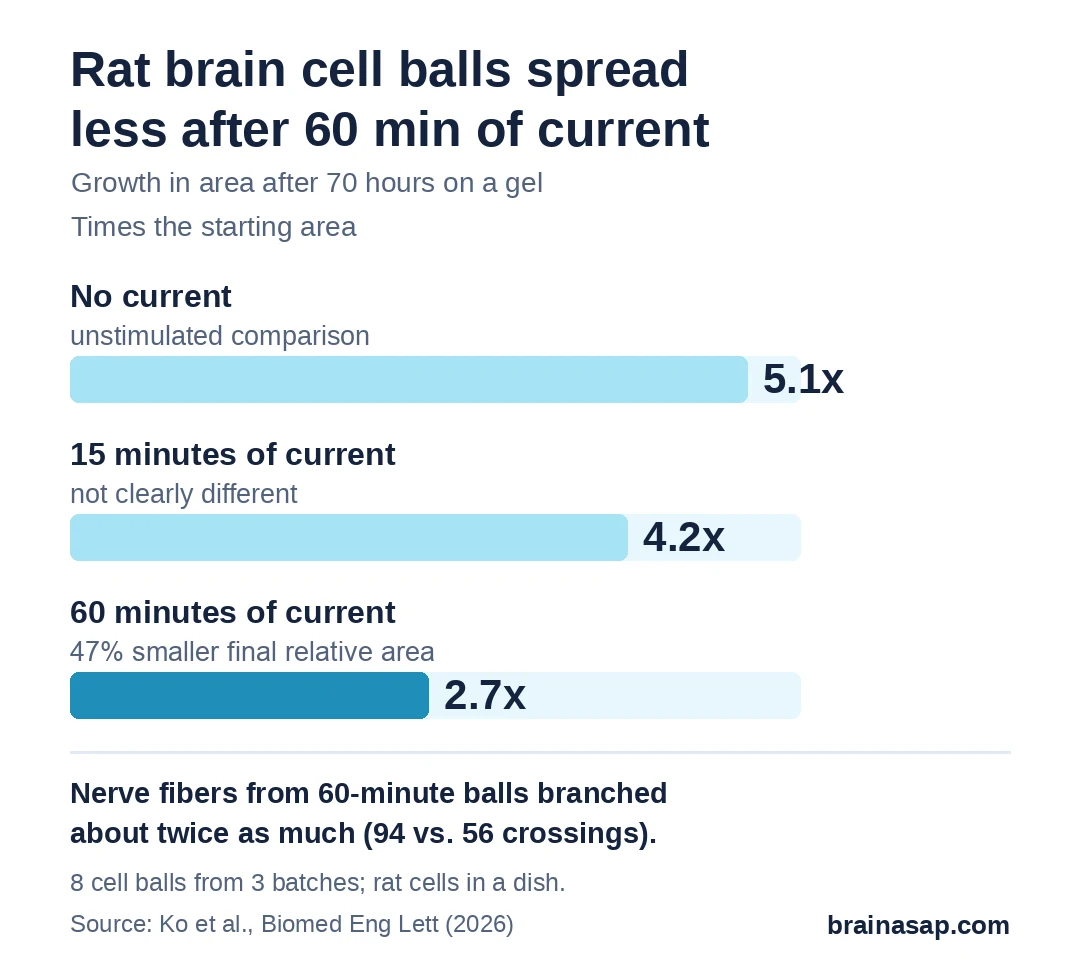
- Less spreading after 60 minutes of current: unstimulated cell balls grew to 5.1 times their starting area in 70 hours; those stimulated for 60 minutes reached only 2.7 times, a 47% smaller final area relative to the starting area.
- 15 minutes didn’t clearly matter: those cell balls grew to 4.2 times their area, not clearly different from unstimulated ones.
- More nerve-fiber branching: far from the ball’s edge, 60-minute balls had about twice as many fiber branches (94 vs. 56 crossings on a counting grid).
- Learning-related genes switched on: Arc, a gene active when brain connections strengthen, rose 2.2-fold after 60 minutes.
- A sticky protein acted as a brake: blocking N-cadherin, which holds cells together, let stimulated balls spread normally again.
Source: Biomedical Engineering Letters (2026) | Ko et al.
Doctors already use electricity on the brain, from deep brain stimulation for Parkinson’s to weak currents through the scalp being tested for memory and mood. What the current actually does to brain tissue, cell by cell, is far less clear.
A 2026 study in Biomedical Engineering Letters, by engineers at KAIST in South Korea, looked at that question in miniature: balls of brain cells about the size of a grain of sand.
Tiny Balls of Rat Brain Cells
The team took cells from the outer layer of young rats’ brains and let them clump together for 7 days into cell balls, called spheroids. Each ball held 3 main brain cell types: neurons, support cells called astrocytes, and immune cells called microglia.
Cell balls were placed on a soft gel and exposed to a steady electric current, at a strength meant to mimic brain stimulation, for:
- 0 minutes (unstimulated comparison)
- 15 minutes
- 60 minutes
A camera then tracked each ball for 72 hours as cells crawled outward onto the gel.
Did the Current Slow Cells Down?
The result? After 60 minutes, yes. For the first 10 hours all groups spread at a similar pace. From about 20 hours on, the 60-minute balls fell behind.
Cells in the 60-minute group also moved more slowly at their peak (16 vs. 17.5 micrometers an hour) and wandered in curved, disorganized paths near the edge instead of heading straight outward.
Oddly, these cells pulled harder on the gel beneath them, especially at the edge, where microglia gathered. Ko and colleagues suggested that this force anchored the ball in place rather than moving it forward.
Fewer Cells Moving, More Nerve Fibers Growing
While the ball as a whole spread less, its neurons did the opposite. On a separate glass surface, nerve fibers from 60-minute balls reached far beyond the dense core and branched heavily.
To count branching, researchers drew rings around each ball and tallied how many fibers crossed them. Beyond 200 micrometers from the edge:
- 60-minute balls: 94 crossings
- Unstimulated balls: 56 crossings
- 15-minute balls: 48 crossings
Genes that switch on when brain connections strengthen also rose. Arc climbed 2.2-fold after 60 minutes, and c-Fos, a fast-acting activity gene, peaked at 5.5-fold after 15 minutes.
A Sticky Protein Acting Like a Brake
N-cadherin is a protein that glues neighboring brain cells together. In unstimulated balls it formed a neat ring near the edge. After 60 minutes of current, it bunched into dense, patchy clusters through the core, with gene activity up 1.66-fold.
To test whether that glue was holding the ball back, researchers added ADH-1, a drug that blocks N-cadherin. It had little effect on unstimulated balls, but stimulated balls then spread like unstimulated ones by 70 hours, with straight, outward paths.
A Dish Is Not a Brain
- Rat cells, not people: taken from young rats and grown for a week.
- No blood vessels: cell balls lack the vessels and full layered structure of real brain tissue.
- Few samples: 8 cell balls from 3 batches for the main spreading results.
- One current type: steady direct current at one strength, unlike the pulsed signals of many brain stimulation devices.
- Microglia role untested: the team called their link to the braking force suggestive, not proven.
Does Longer Stimulation Favor Connections Over Movement?
These findings suggest that how long a current runs may change what brain tissue does with it, holding cells in place while letting nerve fibers grow.
Whether the same shift happens in a living brain, and whether it helps or hurts recovery after injury, is the question this cell work can’t answer yet.
Citation: DOI: 10.1007/s13534-026-00586-9. Ko E, Park E, Shin JH. Electrical stimulation promotes structural plasticity in cortical spheroids by modulating N-cadherin-dependent collective migration and mechanotransduction. Biomed Eng Lett. 2026;16:1273–1285.
Study Design: In vitro experiment on 3D spheroids of postnatal rat cortical cells exposed to DC electrical stimulation (2.0 V/cm) for 0, 15 or 60 minutes.
Sample Size: 8 spheroids from 3 independent cell preparations for spreading and migration; other measures 7 to 17 samples.
Key Statistic: Spreading area at 70 hours 5.1-fold (control) vs. 2.7-fold (60 minutes), a 47% reduction.
Caveat: Rat cells in a dish; no vasculature; small sample; single DC stimulation setting.