TL;DR: In 52 healthy adults, how strongly a tiny brainstem alertness hub responded during waking tasks was linked to the intensity of their REM sleep measured in the lab. The direction of the link depended on the task and on age.
Key Findings
- 52 adults: 34 younger (about 22) and 18 older (about 61).
- Visual focus task: Stronger hub response went with more intense REM sleep and a larger pre-REM brain-wave burst.
- Sound task: Link with REM intensity flipped to negative in older adults.
- REM amount and timing: Not linked to hub activity.
Source: Journal of Biomedical Science (2025) | Mortazavi et al.
Deep in the brainstem sits the locus coeruleus, a small cluster of cells that releases noradrenaline and helps set how alert we are. In animals, it must go quiet for REM (dreaming) sleep to happen. Whether its daytime activity shapes human sleep has been hard to test, because it is so small.
A team at the University of Liège in Belgium used an ultra-powerful 7-Tesla MRI scanner to take a look.
Scanning a Tiny Alertness Hub
The researchers studied 34 younger (about 22 years old) and 18 older (about 61) healthy adults. Each did two tasks in the scanner:
- Visual focus task: Watching an image that flips between two interpretations, which keeps the hub steadily engaged.
- Sound task: Picking out rare target sounds among standard tones, which triggers brief bursts of hub activity.
Participants also slept a night in the lab with EEG to measure REM sleep: how much, how soon it started, how intense its theta brain waves were, and a burst of "sigma" waves just before each REM episode that animal studies tie to the hub.
For younger adults, the brain scan followed the sleep night, about three hours after waking. In older adults, sleep and brain scans were separated by an average of 15.5 months.
A Busier Hub Went With More Intense REM Sleep
For the visual task, a stronger hub response was linked to more intense REM theta waves and to more sigma power just before REM. These links held after correcting for multiple tests.
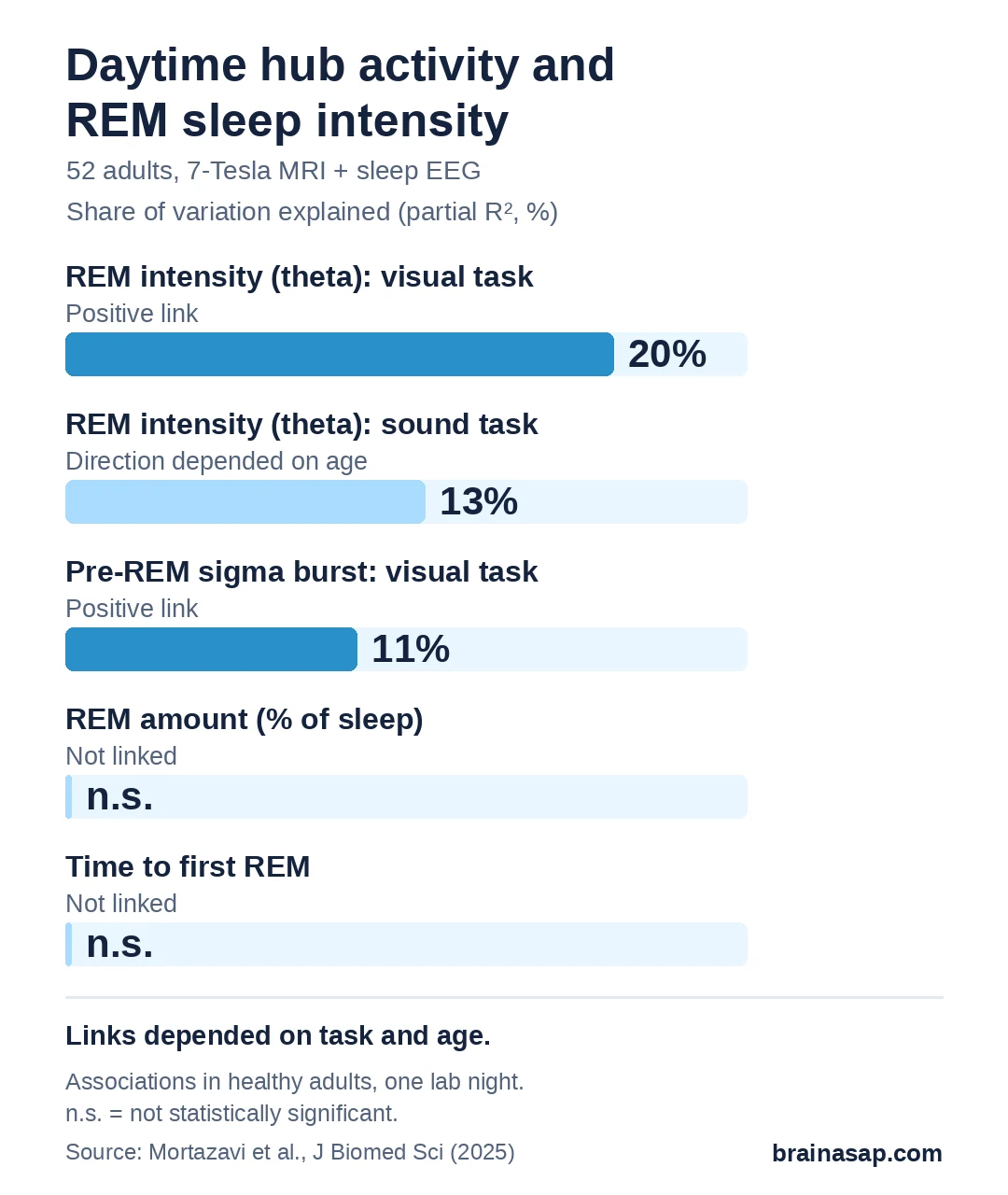
For the sound task, the story was different. The link between hub activity and REM intensity depended on age, and in older adults it turned negative: more hub activity went with less intense REM sleep.
A "Balanced" Hub May Favor Good REM Sleep
The authors suggest the two tasks tap different modes of hub activity, a steady background level and quick bursts. Their interpretation is that good REM sleep needs the steady background level to be balanced, not simply high or low. How much REM people got, and how soon it started, were not linked to hub activity.
Limits of a One-Night Snapshot
- Associations, not cause: The study cannot show the hub controls REM sleep in people.
- Small groups: Especially the 18 older adults, whose sleep and brain scans were also about 15 months apart.
- Indirect measures: MRI signals from such a tiny region are hard to isolate.
- Healthy volunteers: Not people with insomnia or neurological disease.
Relevance for Aging and Brain Disease
The locus coeruleus is one of the first areas affected in Alzheimer's and Parkinson's disease, and sleep complaints rise with age. The open question is whether changes in this hub help explain why REM sleep deteriorates in these conditions, and whether that could be tracked or treated.
Citation: DOI: 10.1186/s12929-025-01127-9. Mortazavi N, Talwar P, Koshmanova E, et al., Vandewalle G. REM sleep quality is associated with balanced tonic activity of the locus coeruleus during wakefulness. J Biomed Sci. 2025;32:35.
Study Design: Cross-sectional study combining 7-Tesla fMRI of the locus coeruleus with in-lab overnight sleep EEG.
Sample Size: 52 healthy adults (34 younger, 18 older).
Key Statistic: Visual-task LC activity and REM theta energy: F(1,45) = 11.39, P = 0.001, partial R² = 0.20.
Caveat: Associations only; small older group; task- and age-dependent results.