TL;DR: Researchers used brain scans to work out a personal target spot for magnetic brain stimulation in each person with schizophrenia. Among 24 patients with an identifiable target, a shorter estimated distance from the standard treatment site was linked to a bigger drop in symptoms such as low motivation. A controlled trial would be needed to test whether personalized targeting improves outcomes.
Key Findings
- A personal target for each patient: the researchers started with scans from 532 people with schizophrenia and 526 without, then used a computer model to estimate targets for patients with a suitable scan pattern.
- Close to, but not on, the standard spot: personal targets sat about 6 mm on average from the spot clinics usually use.
- Closer meant more improvement: among the 24 patients in the distance analysis, a shorter estimated distance was linked to a larger drop in negative symptoms, such as withdrawal and low motivation.
- Brain wiring shifted: after 4 weeks of treatment, measured connectivity changes agreed more closely with the model’s predictions when the estimated distance was smaller.
Source: NeuroImage: Clinical (2026) | Tang, Luo and colleagues
Repetitive transcranial magnetic stimulation, or rTMS, uses a magnetic coil held against the head to send pulses into the brain. It is approved for depression and is being tested in schizophrenia, especially for hard-to-treat symptoms such as low motivation, flat emotion and social withdrawal.
Results in schizophrenia have been mixed. One suspected reason: clinics usually aim the coil at the same spot on everyone's scalp, even though every brain is wired a little differently.
Finding Each Patient's Personal Target
A 2026 study in NeuroImage: Clinical, led by Yingjie Tang and Cheng Luo at the University of Electronic Science and Technology of China in Chengdu, built a computer method to pick a personal stimulation spot for each patient.
It works in 3 steps:
- Find areas where a patient's brain has lost the most tissue compared with healthy people, then work out which brain hub those areas are most wired to. Tang and colleagues called these hubs "epicenters."
- Look within the left dorsolateral prefrontal cortex, a region near the upper front of the head that clinics already stimulate, for the points most strongly connected to those hubs.
- Combine those points into one personal target.
Researchers developed the method on MRI scans from 5 sites, covering 532 people with schizophrenia and 526 without.
Where Did the Hubs Show Up?
Hubs tended to sit in brain networks that handle emotion, attention and planning, plus deep structures such as the thalamus and putamen. Areas that handle vision and movement were rarely involved.
Personal targets were spread out across the stimulated region. On average they sat about 6 mm from the standard spot, a point on the scalp known as F3.
Testing It on Patients Already Treated
Next, the team looked back at 27 inpatients with schizophrenia at a Chengdu hospital who had received rTMS at the standard F3 spot: 20 sessions over 4 weeks. 24 of them had an identifiable hub, so they could be given a personal target after the fact.
A key question: did patients with a shorter estimated distance between the standard spot and their personal target improve more?
They did, moderately. A shorter distance went with a larger drop in negative symptoms (correlation of -0.42, where -1 would be a perfect link). It also went with brain connection changes that better matched what the method predicted (correlation of -0.45).
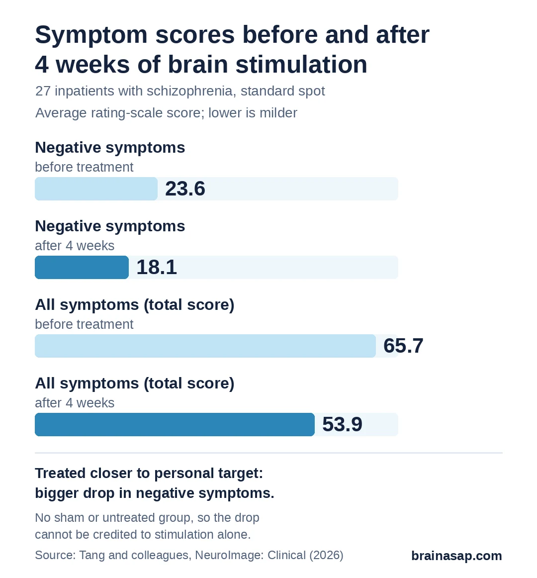
As a group, patients' average negative-symptom score fell from about 23.6 to 18.1 over the 4 weeks on a standard schizophrenia rating scale. With no comparison group, though, that drop can't be credited to the stimulation alone.
Why This Is Only a First Step
- Looking back, not a trial: no patient was treated at a personal target, so the study can't show that personal targeting works better
- Coil position was estimated: actual coil placement wasn't recorded, so the standard spot was approximated for each patient
- Small treatment group: 24 analyzed patients, and the links with improvement were moderate
- No sham group: some improvement could reflect time, medication or hospital care
Tang and colleagues described exactly the trial needed next: patients randomly assigned to personal targets or the standard spot, with everything else matched and coil position tracked at every session.
A Testable Idea for Better-Aimed Treatment
This work offers a way to choose a stimulation spot from each patient's own brain scans, and early data hint that hitting closer to that spot might matter for negative symptoms, which current medicines often don't help much.
Whether aiming at a personal target actually beats the standard spot is the question a randomized trial now has to answer.
Citation: DOI: 10.1016/j.nicl.2026.104055. Tang Y, Jiang S, Huang H, Sun X, Mu C, Duan M, Rodríguez-Labrada R, Yao D, Luo C. An Epicenter-guided computational framework for personalized target of repetitive transcranial magnetic stimulation in schizophrenia. NeuroImage: Clinical. 2026;52:104055. PMID: 42710367.
Study Design: Computational framework development on multisite structural and resting-state MRI (gray-matter deviation epicenters mapped to normative connectivity, left DLPFC peak-connectivity voxels fused by harmonic centrality); retrospective validation in an open-label rTMS cohort (10 Hz, 110% resting motor threshold, 20 sessions over 4 weeks, F3 placement) with activity-flow modeling.
Sample Size: Discovery: 532 people with schizophrenia and 526 healthy controls (5 sites). Treatment cohort: 27 inpatients (24 with identifiable epicenters) and 88 same-scanner healthy controls.
Key Statistic: Personalized target to F3 distance 6.23 ± 2.30 mm (discovery), 5.82 ± 2.00 mm (treatment cohort); distance vs negative-symptom improvement Spearman r = -0.42, p = 0.04; distance vs predicted-observed connectivity change similarity r = -0.45, p = 0.03.
Caveat: Retrospective analysis; no patient stimulated at a personalized target; actual coil coordinates not recorded; no sham or untreated control; small treatment cohort.