
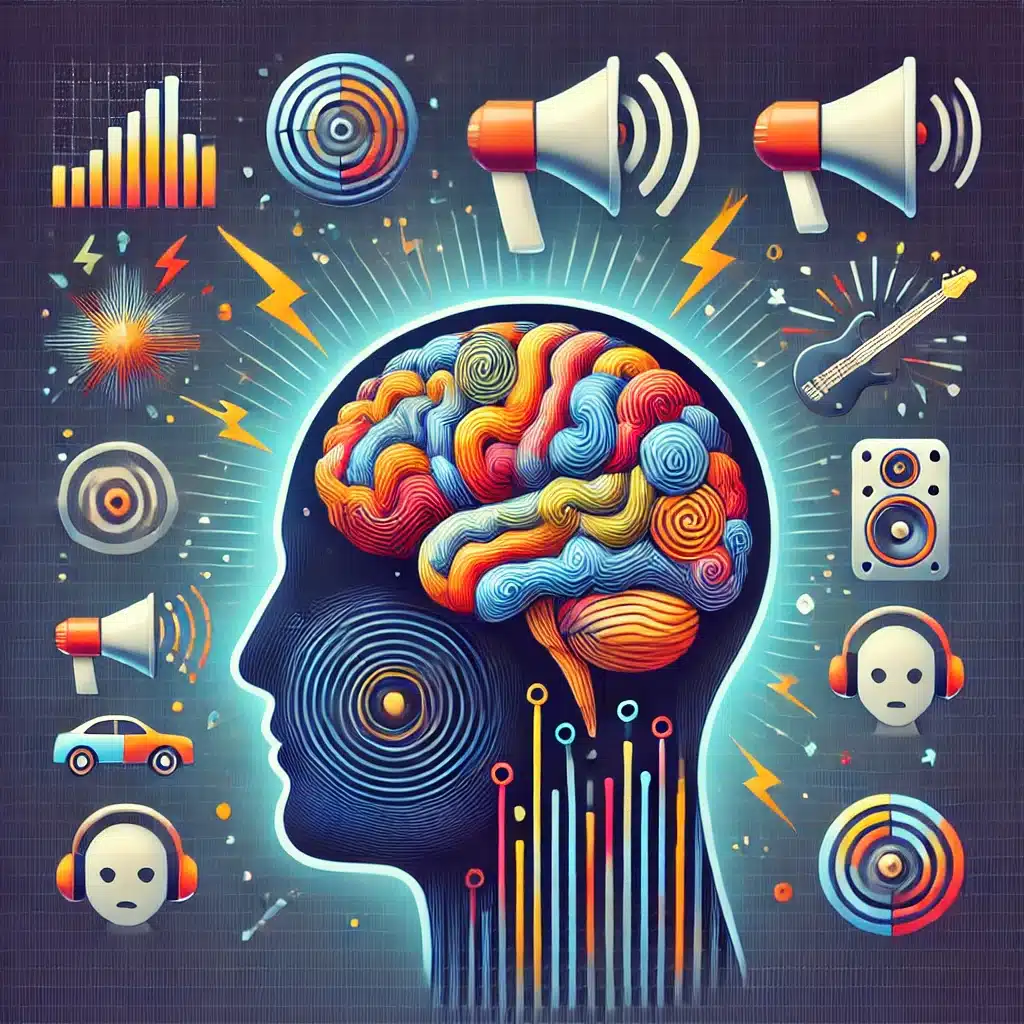
TLDR: Patients with noise-induced hearing loss (NIHL) exhibit significant changes in the white matter (WM) microstructure of their brains, particularly affecting the auditory and language pathways.
Highlights:
- A study compared 122 patients with varying degrees of NIHL and 84 healthy controls.
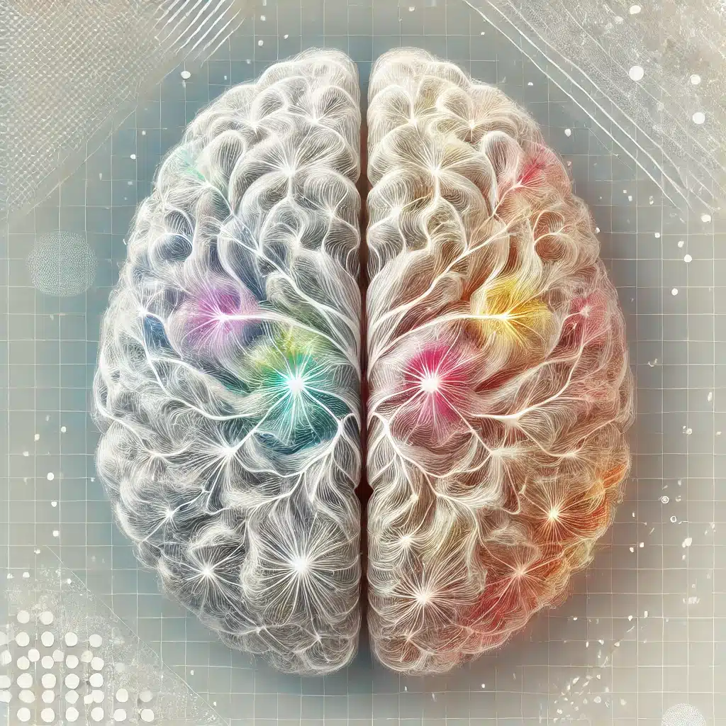
- Diffusion tensor imaging (DTI) revealed significant differences in the white matter microstructure, particularly in regions associated with auditory and language processing, such as the left inferior longitudinal fasciculus (ILF) and left inferior fronto-occipital fasciculus (IFOF).
- There was a significant negative correlation between the white matter integrity and anxiety levels in patients with NIHL.
- Longer noise exposure was correlated with higher hearing thresholds and greater structural brain changes.
- Patients with NIHL showed lower fractional anisotropy (FA), indicating reduced white matter integrity in several brain regions compared to healthy controls.
Source: Brain & Behavior (2024)
Main Findings of the Study on Noise-Induced Hearing Loss (NIHL)
1. Participants with NIHL
The study involved 122 patients with noise-induced hearing loss (NIHL) and 84 healthy controls.
Patients were categorized into three groups: mild NIHL (MP), relatively severe NIHL (RSP), and undetermined severity.
2. White Matter Microstructure Changes
Affected Regions: Significant changes were found in the white matter (WM) of the brain, specifically in the left inferior longitudinal fasciculus (ILF) and left inferior fronto-occipital fasciculus (IFOF).
Broader Impact: These changes affect areas important for auditory and language processing, indicating that long-term noise exposure can alter brain structures related to hearing and language.
3. Correlation with Anxiety
Anxiety Scores: The study found a significant negative correlation between WM integrity and anxiety levels. Patients with more significant WM changes tended to have higher anxiety scores.
Hearing Loss & Anxiety: Anxiety levels were higher in patients with NIHL compared to healthy controls, suggesting a link between hearing loss and mental health.
4. Noise Exposure Effects
Hearing Threshold: Longer noise exposure times were associated with higher hearing thresholds, meaning greater hearing loss.
Brain Structure: Extended noise exposure also correlated with more pronounced changes in brain structure, reinforcing the impact of prolonged noise on brain health.
5. White Matter Integrity
Patients with NIHL had lower fractional anisotropy (FA) values in several brain regions, indicating reduced white matter integrity.
This was observed in areas like the bilateral corticospinal tract (CST) and right fronto-pontine tract (FPT).
How Noise-Induced Hearing Loss May Lead to White Matter Changes in the Brain (Mechanisms)
1. Chronic Noise Exposure
Prolonged Noise Damage: Continuous exposure to high levels of noise can lead to sustained stress on the auditory system, causing degeneration of the sensory hair cells and spiral ganglion neurons in the inner ear.
Neural Pathway Strain: The damage to the inner ear may strain the neural pathways that process sound, leading to compensatory changes in the brain’s white matter to adapt to the reduced auditory input.
2. Auditory Deprivation
Reduced Auditory Input: With the loss of hearing, the brain receives less auditory information, leading to decreased stimulation of the auditory pathways.
Neuroplasticity: The brain may undergo neuroplastic changes, where it reorganizes itself to compensate for the lack of auditory input. This can result in alterations in the white matter structure as the brain adapts to new ways of processing information.
3. Stress & Anxiety
Emotional Impact: Hearing loss can lead to increased anxiety and stress, which are known to affect brain structure and function.
Physiological Stress Responses: Chronic stress and anxiety can lead to changes in the brain’s white matter due to the release of stress hormones, which can affect neural connectivity and integrity.
Possible Effects of White Matter Microstructure Changes from NIHL
1. Cognitive Function
Language & Communication: Changes in white matter in regions such as the ILF and IFOF, which are involved in language processing, can lead to difficulties in understanding and producing speech.
Memory & Learning: The cingulum (hippocampus) is crucial for memory and learning. Alterations in this area can impact these cognitive functions, potentially leading to difficulties in recalling information and learning new skills.
2. Emotional & Mental Health
Increased Anxiety: Structural changes in the white matter associated with anxiety (such as in the right fronto-pontine tract) can exacerbate feelings of anxiety, leading to a vicious cycle of worsening mental health.
Emotional Regulation: The cingulum and other affected regions are involved in emotional regulation. Changes in these areas can lead to difficulties in managing emotions, resulting in increased irritability, mood swings, and other emotional disturbances.
3. Auditory Processing
Sound Localization and Processing: Damage to the white matter pathways that process auditory information can affect the brain’s ability to localize and interpret sounds. This can lead to difficulties in understanding speech, especially in noisy environments.
Tinnitus: Changes in the auditory pathways can contribute to the perception of phantom sounds or tinnitus, which is a common and distressing symptom in individuals with hearing loss.
4. Quality of Life
Social Isolation: Difficulties in communication and increased anxiety can lead to social withdrawal and isolation, reducing the overall quality of life.
Functional Impairments: Cognitive and emotional challenges can affect daily functioning, making it harder for individuals to perform tasks that require concentration, memory, and emotional stability.
Study Details: Hearing Loss & White Matter Microstructure (2024)
Participants
Total Enrollment: 122 patients with noise-induced hearing loss (NIHL) and 84 healthy controls.
Group Categorization: Patients were divided into three groups based on the severity of their hearing loss:
- Mild Patients (MP): 71 participants.
- Relatively Severe Patients (RSP): 28 participants.
- Undetermined Severity: 11 participants (lost to follow-up).
Healthy Controls (HCs): 75 participants after data processing and elimination.
Methods
1. Clinical Data Collection
Demographics: Age and education level.
Hearing Assessment: Hearing threshold measurements.
Noise Exposure: Occupational noise exposure time.
Mental Health Evaluations:
- Mini-Mental State Examination (MMSE) for cognitive status.
- Tinnitus Handicap Inventory (THI) for tinnitus severity.
- Hamilton Anxiety Scale (HAMA) for anxiety levels.
2. MRI & Diffusion Tensor Imaging (DTI)
MRI Scanning: Performed using a GE Discovery MR 750 3.0 T scanner.
- T1WI3DFSPGR sequence for brain structure imaging.
- DTI with echo planar imaging sequence for white matter analysis.
DTI Analysis: Utilized tract-based spatial statistics (TBSS) and region of interest (ROI) analysis to assess differences in DTI parameters (fractional anisotropy [FA], axial diffusivity [AD], mean diffusivity [MD], and radial diffusivity [RD]) among the groups.
3. Statistical Analysis
ANOVA & t-tests: Used to analyze differences in clinical data, hearing thresholds, noise exposure times, and DTI parameters.
Pearson Correlation: Assessed correlations between clinical data (e.g., noise exposure time, hearing threshold) and DTI parameters.
Multiple Comparisons: Bonferroni correction was applied for multiple group comparisons to ensure statistical significance.
Limitations
- Sample Size: The RSP group had a smaller sample size, which may limit the generalizability of the findings for this subgroup.
- Noise Exposure Assessment: The study did not account for other sources of noise exposure, such as personal music players, which could influence hearing loss and brain changes.
- Neuropsychological Testing: The study included few neuropsychological tests. Future research should incorporate specific cognitive and emotional function tests related to NIHL.
- Tinnitus Evaluation: The study faced challenges in evaluating tinnitus incidence, variation, and severity, which are important factors in understanding NIHL.
- Spatial Resolution: The spatially coarse feature of diffusion imaging may affect the ability to detect fine-grained NIHL-related changes, particularly in the auditory cortex.
Strategies to Mitigate the White Matter Microstructure Changes in Hearing Loss
1. Early Intervention & Prevention
Noise Protection: Use of protective gear such as earplugs or noise-canceling headphones in noisy environments to prevent further damage.
Workplace Regulations: Implementing and enforcing stricter noise regulations in occupational settings to reduce exposure to harmful noise levels.
2. Hearing Aids & Cochlear Implants
Hearing Aids: Providing hearing aids to amplify sound can help reduce the strain on the auditory system and improve hearing function.
Cochlear Implants: For severe cases, cochlear implants can bypass damaged parts of the ear and directly stimulate the auditory nerve, restoring some level of hearing.
3. Cognitive & Behavioral Therapies
Cognitive Behavioral Therapy (CBT): CBT can help manage anxiety and stress, which are linked to white matter changes, by teaching coping mechanisms and reducing negative thought patterns.
Tinnitus Retraining Therapy (TRT): This therapy can help individuals habituate to the tinnitus sounds, reducing their impact on daily life.
4. Auditory Training Programs
Listening Exercises: Engaging in exercises designed to improve auditory processing and speech comprehension can stimulate the auditory pathways and potentially counteract some of the structural changes.
Music Therapy: Music therapy has been shown to enhance neural plasticity and improve auditory processing skills.
5. Pharmacological Interventions
Anti-Anxiety Medications: Prescribing medications to manage anxiety can help reduce the impact of stress on the brain’s white matter.
Neuroprotective Drugs: Research into drugs that protect against neural damage or promote neural repair could offer future treatment options.
6. Lifestyle Modifications
Healthy Diet: A diet rich in antioxidants and omega-3 fatty acids can support brain health and reduce inflammation.
Regular Exercise: Physical activity is known to promote neurogenesis and improve overall brain function, potentially mitigating some of the changes in white matter.
7. Technological Innovations
Brain Stimulation Techniques: Techniques like transcranial magnetic stimulation (TMS) may enhance neural plasticity and improve auditory and cognitive functions.
AI & Machine Learning: Developing AI-driven hearing aids that adapt to the user’s environment and optimize sound processing in real-time.
Conclusion: Noise Induced Hearing Loss & White Matter Changes
This study provides critical insights into how noise-induced hearing loss (NIHL) affects brain structure, particularly the white matter microstructure.
Significant changes were observed in key regions associated with auditory and language processing, indicating that prolonged noise exposure leads to structural brain alterations.
These changes were also correlated with increased anxiety levels, highlighting the broader impact of NIHL on mental health.
Early intervention, such as using protective gear and providing auditory training, along with advanced therapies like cognitive-behavioral therapy and potential pharmacological treatments, can help mitigate these effects.
Despite some limitations, such as sample size and the scope of neuropsychological tests, the findings underscore the importance of addressing NIHL to preserve cognitive function and overall quality of life.
Future research should focus on refining these strategies and exploring additional preventive measures to protect individuals from the adverse effects of prolonged noise exposure.
References
- Study: Alterations of the cerebral microstructure in patients with noise-induced hearing loss: A diffusion tensor imaging study (2024)
- Authors: Ranran Huang et al.


