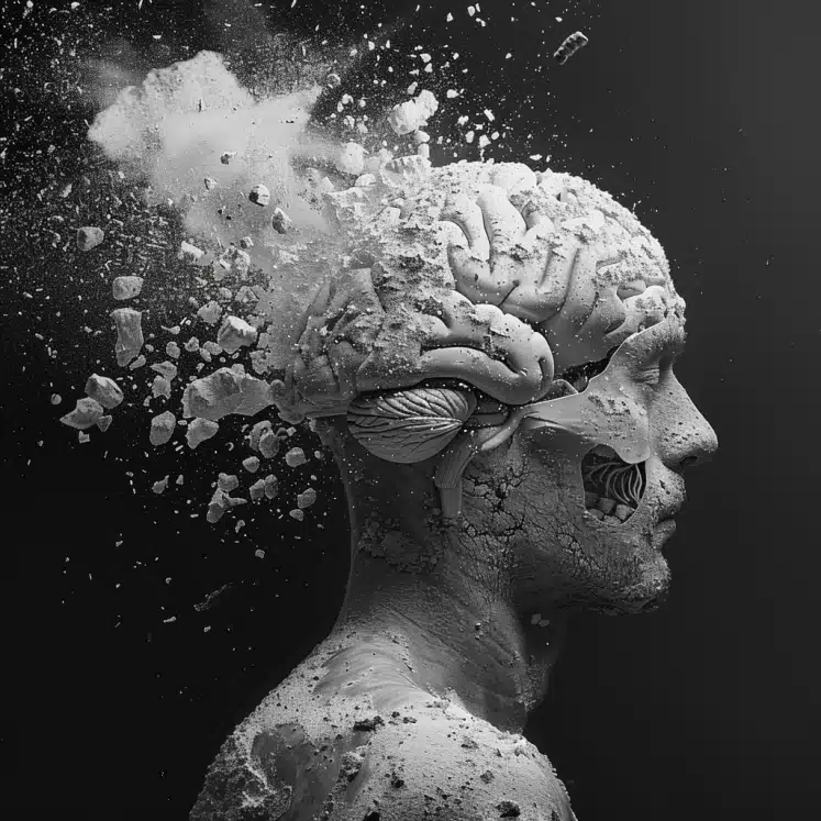
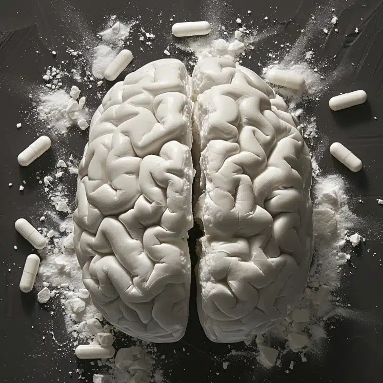
Cocaine Use Disorder (CUD) patients exhibit widespread gray matter atrophy, with specific brain volume deficits associated with crack-cocaine use and relapse outcomes.
Highlights:
- Widespread Gray Matter Atrophy: CUD patients show significant gray matter volume reductions in frontal and temporal cortices, cerebellum, and subcortical structures compared to healthy controls.
- Route of Use: Crack-cocaine use is linked to specific volume deficits in the right inferior and middle temporal gyri and the right fusiform gyrus.
- Relapse Prediction: Smaller volumes in the cerebellar vermis at detoxification are associated with relapse within three months.
- Cortical & Subcortical Damage: Structural damage is observed in both cortical areas, including the insula and various parts of the limbic system, and subcortical regions such as the thalamus and striatum.
- Socioeconomic Factors: Crack-cocaine users tend to have more severe socio-economic disadvantages and worse treatment outcomes than those using cocaine hydrochloride.
Source: Psychiatry Research: Neuroimaging (2024)
Major Findings: Cocaine Addiction & Brain Damage (2024)
1. Widespread Gray Matter Atrophy in Cocaine Patients
Patients with Cocaine Use Disorder (CUD) have significant reductions in gray matter volume across several brain regions.
The most affected areas include the frontal and temporal cortices, which are critical for decision-making, memory, and emotional regulation.
The study also found shrinkage in the cerebellum, which plays a role in motor control and cognitive functions, and in subcortical structures like the thalamus and striatum, which are involved in sensory processing and reward pathways.
2. Specific Brain Volume Deficits Associated with Crack-Cocaine Use
Patients who primarily used crack-cocaine exhibited specific reductions in brain volume that differed from those who used cocaine hydrochloride.
Crack-cocaine users had lower volumes in the right inferior and middle temporal gyri and the right fusiform gyrus.
These regions are crucial for processing sensory information and facial recognition, indicating that crack-cocaine might have more severe effects on these cognitive functions compared to other forms of cocaine.
3. Relapse Prediction Based on Cerebellar Vermis Volume
The size of the cerebellar vermis can predict whether a CUD patient will relapse within three months after detoxification.
Patients who relapsed had smaller volumes in the cerebellar vermis at the start of their detox program compared to those who remained abstinent.
The cerebellar vermis is involved in balance and coordination, but it also plays a role in emotional regulation and cognitive processing.
This suggests that reduced volume in this area might be linked to a higher vulnerability to relapse.
4. Cortical & Subcortical Brain Damage
Chronic cocaine use leads to structural damage in both cortical and subcortical brain regions.
Damage was particularly notable in the insula, which is involved in consciousness and homeostasis, and the limbic system, including the amygdala and hypothalamus, which are critical for emotional responses and hormone regulation.
The striatum, part of the brain’s reward system, also showed volume reductions, highlighting the extensive impact of cocaine on brain function.
5. Differences Between Crack-Cocaine & Cocaine HCL Users
Crack-cocaine users generally experience more severe socio-economic challenges and worse treatment outcomes compared to those using cocaine hydrochloride.
Crack-cocaine users were found to be older, with more unstable housing situations and lower employment rates.
These socio-economic factors contribute to more significant cognitive impairments and a higher likelihood of relapse, underscoring the need for tailored treatment approaches that address both medical and socio-economic issues.
Study Overview: Brain Changes in Cocaine Use Disorder (CUD)
The study aimed to investigate the structural brain abnormalities in patients with Cocaine Use Disorder (CUD), examining the impact of different routes of cocaine administration (crack-cocaine vs. cocaine hydrochloride) and their predictive value for relapse.
Sample
- Participants: 55 patients with severe CUD (26 using crack-cocaine and 29 using cocaine hydrochloride) entering inpatient detoxification, and 38 matched healthy controls.
- Follow-up: A 3-month outpatient follow-up to determine treatment outcomes (relapse vs. abstinence).
Methods
- Study Design: Anatomical MRI study with a Voxel-Based Morphometry (VBM) approach.
- Data Collection: MRI scans were conducted within one week of detoxification for CUD patients. Sociodemographic and clinical data, including substance use patterns, were collected at baseline and during follow-up interviews.
- Analysis: Two-sample t-tests comparing brain volumes between CUD patients and healthy controls, between crack-cocaine and cocaine hydrochloride users, and between relapsers and abstainers.
Limitations
- Comorbid Conditions: The CUD patient group had various comorbid conditions and medications that were not controlled, potentially biasing results.
- Sample Size: The group of abstainers was small (9 patients), which might have affected the statistical power of the analysis regarding relapse.
- Generalizability: The study’s findings may not be generalizable to all CUD patients due to the specific demographic and clinical characteristics of the sample.
- Poly-Substance Use: Many patients were poly-substance users, which could confound the specific effects of cocaine on brain structure.
Potential Reasons for Brain Abnormalities in Cocaine Use Disorder (CUD) Patients

1. Neurotoxic Effects of Cocaine
Mechanism: Cocaine is known to increase dopamine levels by blocking the dopamine transporter. Chronic cocaine use leads to neurotoxicity due to excessive dopamine levels, oxidative stress, and excitotoxicity.
Impact: This results in neuronal damage and loss, particularly in regions involved in reward processing, decision-making, and emotional regulation.
2. Specific Brain Regions Affected
Frontal Cortex
- Role in Healthy Brains: The frontal cortex, including the prefrontal cortex, is critical for executive functions, decision-making, and impulse control.
- Cocaine Impact: Reduced gray matter volume in the frontal cortex of CUD patients impairs these functions, leading to poor decision-making and increased impulsivity.
Temporal Cortex
- Role in Healthy Brains: The temporal cortex is involved in memory processing, auditory perception, and language.
- Cocaine Impact: Decreased volume in the temporal cortex can result in memory deficits and difficulties in processing sensory information.
Cerebellum
- Role in Healthy Brains: The cerebellum regulates motor control and coordination and contributes to cognitive functions.
- Cocaine Impact: Shrinkage in the cerebellum, particularly in the vermis, can lead to motor coordination problems and cognitive impairments.
Limbic System (including Amygdala and Hypothalamus)
- Role in Healthy Brains: The limbic system is essential for emotion regulation, motivation, and memory.
- Cocaine Impact: Damage to these areas can cause emotional instability, heightened stress responses, and motivational deficits.
Striatum
- Role in Healthy Brains: The striatum is part of the brain’s reward system, involved in reinforcing behaviors and habit formation.
- Cocaine Impact: Reduced volume in the striatum can diminish the ability to experience pleasure from normal activities, leading to increased drug-seeking behavior.
Insula
- Role in Healthy Brains: The insula is involved in interoceptive awareness, emotional processing, and decision-making.
- Cocaine Impact: Atrophy in the insula can affect how individuals process emotions and make decisions, contributing to continued substance use despite negative consequences.
3. Comparison to Healthy Brains
Volume Differences: In healthy brains, these regions maintain normal gray matter volume, which supports proper cognitive, emotional, and motor functions.
Functional Integrity: Healthy individuals exhibit robust connectivity and functionality in these areas, leading to better impulse control, emotional regulation, memory, and decision-making.
Neuroplasticity: Healthy brains have greater neuroplasticity, allowing for better recovery and adaptation to changes, unlike the damaged neural structures in CUD patients.
Conclusion: Brain Structure in Cocaine Use Disorder
The study reveals significant brain structural abnormalities in patients with Cocaine Use Disorder (CUD), highlighting the extensive impact of chronic cocaine use on both cortical and subcortical regions.
Specific deficits were found to be associated with the route of administration, with crack-cocaine users exhibiting more severe volume reductions in critical brain areas compared to those using cocaine hydrochloride.
Additionally, smaller volumes in the cerebellar vermis were identified as a predictor for relapse within three months post-detoxification.
These findings underscore the importance of targeted interventions that address both the neurotoxic effects of cocaine and the specific needs of different user groups.
By leveraging neuroplasticity through cognitive rehabilitation, pharmacological treatments, and brain stimulation techniques, there is potential to repair and restore brain function, supporting long-term recovery.
Comprehensive treatment strategies that include behavioral therapies, lifestyle modifications, and strong social support systems are crucial for improving outcomes for individuals with CUD.
References
- Study: Brain alterations in Cocaine Use Disorder: Does the route of use matter and does it relate to the treatment outcome? (2024)
- Authors: Margaux Poireau et al.


