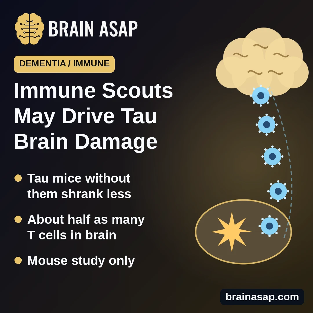
TL;DR: In mice with tau-driven brain damage, removing a type of immune "scout" cell outside the brain cut the number of killer T cells reaching the brain and protected against brain shrinkage. Tau tangles themselves barely changed.
Key Findings
- Less brain damage: Mice lacking the scout cells kept more hippocampus and nearby cortex.
- Fewer T cells in the brain: T cells fell from about 70% to 40% of immune cells entering the brain.
- Calmer support cells: Less activation of microglia and astrocytes, mostly in males.
- Priming happens outside the brain: In lymph nodes that drain the brain.
- Tau buildup: Largely unchanged.
Source: Nature Neuroscience (2026) | Hu et al.
In Alzheimer's disease and related "tauopathies," tangles of a protein called tau build up inside neurons. But tau is not acting alone. Brains with more tau damage also contain more CD8 T cells, the immune system's killer cells, and earlier work from the same team showed that removing T cells protected tau mice.
That raised a question: what switches those T cells on and sends them into the brain? Researchers at Washington University in St. Louis traced it to a specific scout cell in the body's immune system.
Immune Scouts That Train Killer T Cells
Type 1 conventional dendritic cells (cDC1s) are rare immune cells that pick up protein fragments and show them to CD8 T cells, training those T cells to attack. This "cross-presentation" is how the body primes T cells against viruses and tumors.
The team used mice with a human tau mutation and the human APOE4 gene, which develop heavy brain damage by 9.5 months. They compared:
- Tau mice with scouts: Normal dendritic cells.
- Tau mice without scouts: Genetically unable to make cDC1s (or, in other experiments, unable to cross-present).
Without the Scouts, Brains Held Up Better
By 9.5 months, tau mice with normal scouts had lost much of the hippocampus and nearby cortex. Tau mice without scouts showed significant preservation of these regions, more surviving neurons in the hippocampus, and better nest building, a sign of healthier brain function. A blood marker of nerve damage was lower in males.
Microglia and astrocytes, the brain's support and immune cells, were less activated, mainly in male mice. The researchers suggest female mice had less brain damage to begin with.
Fewer Killer T Cells Made It Into the Brain
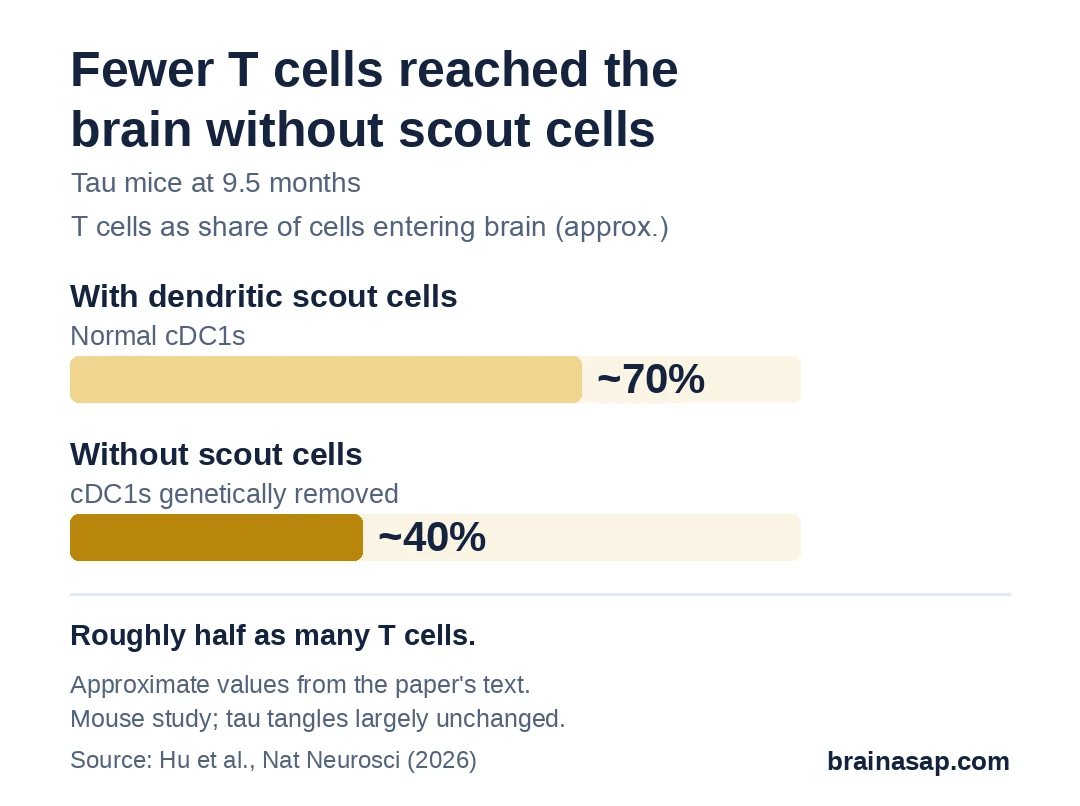
Without the scouts, about 40% fewer immune cells entered the brain overall, and T cells dropped from about 70% to about 40% of them, roughly half as many. The loss was in CD8 killer T cells; helper (CD4) T cells were unchanged. The CD8 T cells that remained showed less clonal expansion, a sign they had not been properly primed.
The Training Happens in Lymph Nodes
Scout cells were barely detectable in the brain itself. Using a tracer protein made in brain neurons, the researchers showed that brain-derived fragments were presented to T cells in lymph nodes, including those draining the brain in the neck. That points to priming outside the brain.
Brain tissue from people who had died with several tauopathies also contained CD8 T cells, consistent with the mouse findings.
Where the Evidence Stops
- Mouse genetics: Removing a cell type from birth is not a treatment.
- Tau itself unchanged: The benefit came without much change in tau tangles.
- Sex differences: Several protections were clearer in males.
- Immune trade-offs: These scouts also fight infections and cancer, so blocking them could carry risks.
A Target Outside the Brain
The study suggests that part of tau-driven brain damage depends on immune training in the body, not in the brain. The open question is whether that pathway can be dialed down safely in people, without weakening protection against infection and cancer.
Citation: DOI: 10.1038/s41593-026-02427-5. Hu H, Lin PBC, Zeng C, et al. Priming of CD8+ T cells by peripheral dendritic cells exacerbates tau-mediated neurodegeneration. Nat Neurosci. 2026.
Study Design: Mouse genetics (P301S tau/APOE4 mice lacking cDC1s or cross-presentation), histology, flow cytometry, single-cell sequencing, antigen tracing; human postmortem tissue.
Sample Size: About 19 to 22 tau mice per sex per group for key brain measures; 8 human tauopathy and 5 control brains.
Key Statistic: Conventional T cells about 70% vs 40% of CD45-high immune cells entering the brain (~50% reduction).
Caveat: Mouse model; tau aggregation not strongly altered; effects varied by sex and measure.