This study shows that type 2 diabetes causes decreased blood flow and changes in neural activity in specific parts of the hippocampus, which may initially compensate for cognitive decline but deteriorate with worsening diabetes.
Highlights:
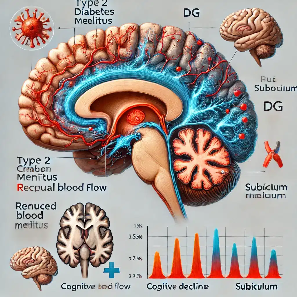
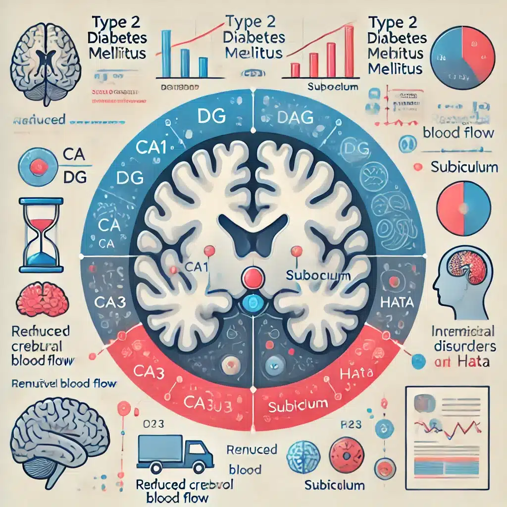
- Decreased Perfusion: Patients with type 2 diabetes have lower blood flow in the right hippocampal CA1, DG, and subiculum areas.
- Increased Neural Activity: The left hippocampal CA3, subiculum, and bilateral HATA regions show heightened neural activity in diabetic patients, potentially as an adaptive response.
- Enhanced Coordination: Diabetic patients exhibit better local neural coordination in the left hippocampal CA3, DG, HATA, and subiculum.
- Chronic Hyperglycemia Impact: The right hippocampal subiculum’s decreased perfusion is linked to long-term high blood sugar levels.
- Compensation Decline: Despite initial compensation, neural activity and coordination diminish as metabolic disorders in diabetes worsen.
Source: Brain & Behavior (2024)
Major Findings: Type 2 Diabetes Linked to Lower Cerebral Blood Flow (CBF) vs. Healthy Controls (2024)
1. Decreased Blood Flow in Specific Hippocampal Regions
Patients with type 2 diabetes (T2DM) showed a significant reduction in blood flow (perfusion) in particular parts of the right hippocampus, specifically:
- Cornu Ammonis Area 1 (CA1)
- Dentate Gyrus (DG)
- Subiculum
These areas are critical for processing and transmitting information in the brain, and reduced blood flow here suggests that diabetes impairs the brain’s ability to function properly.
This reduction in blood flow was linked to chronic high blood sugar levels, a common issue in T2DM.
2. Increased Neural Activity as Compensation
In contrast to the decreased blood flow, the study found increased spontaneous neural activity in the left hippocampus in the following regions:
- Cornu Ammonis Area 3 (CA3)
- Subiculum
- Hippocampus-Amygdala Transition Area (HATA) (on both sides)
This suggests that these regions might be working harder to compensate for the damage or inefficiency caused by diabetes.
This overactivity can be seen as the brain’s way of trying to maintain cognitive functions, such as memory and learning, despite the adverse effects of the disease.
3. Enhanced Local Neural Coordination
In addition to increased neural activity, patients with T2DM exhibited better local coordination among neurons in the left hippocampus, specifically in:
- CA3
- DG
- HATA
- Subiculum
This improved coordination indicates that the brain regions are working together more effectively to process information, potentially as another form of compensation for the damage caused by diabetes.
4. Impact of Chronic Hyperglycemia
The study found a strong link between chronic hyperglycemia (long-term high blood sugar) and decreased perfusion in the right hippocampus’s subiculum region.
High levels of HbA1c, a marker of chronic blood sugar levels, were negatively correlated with blood flow in this area, suggesting that sustained high blood sugar can lead to significant damage and reduced function in the brain.
5. Decline in Compensatory Mechanisms
Despite the initial compensatory increase in neural activity and coordination, these mechanisms seem to decline as metabolic disorders worsen in T2DM.
For example, higher levels of fasting insulin (FINS) and insulin resistance (measured by HOMA-IR) were linked to reduced neural activity and coordination in specific hippocampal regions.
This indicates that as the metabolic control worsens, the brain’s ability to compensate diminishes, potentially leading to further cognitive decline.
Potential Causes of Lower Cerebral Blood Flow (CBF) in Type 2 Diabetes
Type 2 diabetes mellitus (T2DM) is linked to lower cerebral blood flow (CBF) due to several interconnected causes and mechanisms, primarily related to chronic hyperglycemia and insulin resistance.
1. Chronic Hyperglycemia
Vascular Damage: Prolonged high blood sugar levels cause damage to blood vessels, leading to endothelial dysfunction. This impairs the ability of blood vessels to dilate properly, reducing blood flow to the brain.
Polyol Pathway Activation: Chronic hyperglycemia activates the polyol pathway, increasing sorbitol and fructose levels, which can cause osmotic and oxidative stress, further damaging blood vessels.
2. Insulin Resistance
Reduced Nitric Oxide (NO) Production: Insulin resistance reduces the production of NO, a vasodilator crucial for maintaining blood vessel health and proper blood flow. Reduced NO levels lead to vasoconstriction and lower CBF.
Increased Inflammation: Insulin resistance is associated with higher levels of inflammatory cytokines, which contribute to vascular inflammation and stiffness, impairing blood flow.
3. Oxidative Stress
Advanced Glycation End Products (AGEs): Chronic hyperglycemia leads to the formation of AGEs, which accumulate in blood vessels and promote oxidative stress and inflammation, contributing to vascular damage and reduced CBF.
Mitochondrial Dysfunction: High blood sugar and insulin resistance cause mitochondrial dysfunction, leading to increased production of reactive oxygen species (ROS) that damage vascular cells.
4. Diacylglycerol-Protein Kinase C (DAG-PKC) Pathway
Activation of PKC: Hyperglycemia activates the DAG-PKC pathway, which affects the regulation of blood flow by altering the function of various proteins involved in vascular tone and blood flow regulation. PKC activation leads to vasoconstriction and impaired CBF.
5. Microvascular Disease
Capillary Rarefaction: Chronic hyperglycemia and insulin resistance contribute to the loss of capillaries (capillary rarefaction) in the brain, reducing the overall blood supply and leading to lower CBF.
Blood-Brain Barrier (BBB) Disruption: T2DM can disrupt the integrity of the BBB, leading to increased permeability and leakage, which impairs the regulation of blood flow to the brain.
6. Neurovascular Coupling Impairment
Impaired Neurovascular Response: T2DM affects the mechanisms of neurovascular coupling, where neural activity is less effectively matched with corresponding changes in blood flow. This impairment can result in inadequate blood supply to active brain regions.
Potential Effects & Long-Term Consequences of Reduced Cerebral Blood Flow in T2DM
1. Cognitive Decline
- Memory Impairment: Reduced blood flow to the hippocampus, critical for memory formation and recall, can lead to significant memory deficits.
- Executive Dysfunction: Lower CBF can impair functions such as planning, decision-making, and problem-solving, impacting daily activities and quality of life.
- Dementia Risk: Chronic reduced perfusion may increase the risk of developing dementia, including Alzheimer’s disease, due to sustained neuronal damage and degeneration.
2. Mood Disorders
- Depression: Insufficient blood flow to brain regions involved in mood regulation, like the prefrontal cortex, can contribute to the development of depression.
- Anxiety: Reduced CBF may also affect regions associated with anxiety, leading to increased susceptibility to anxiety disorders.
3. Neurological Impairments
- Stroke: Reduced CBF can elevate the risk of stroke by contributing to the development of cerebrovascular disease and promoting clot formation.
- Motor Function Decline: Decreased perfusion in motor-related areas of the brain can impair coordination, balance, and overall motor skills.
Strategies to Reverse Reduced Cerebral Blood Flow in T2DM
1. Blood Sugar Control
Medications: Use antidiabetic medications (e.g., metformin, insulin) to maintain blood glucose levels within a target range.
Diet: Adopt a balanced diet rich in whole grains, vegetables, lean proteins, and healthy fats to stabilize blood sugar levels.
2. Physical Activity
Regular Exercise: Engage in at least 150 minutes of moderate-intensity aerobic exercise per week. Exercise improves insulin sensitivity and promotes vascular health.
Strength Training: Incorporate resistance exercises to enhance overall metabolic health and blood flow.
3. Lifestyle Modifications
Smoking Cessation: Quit smoking to improve vascular health and reduce the risk of cerebrovascular disease.
Alcohol Moderation: Limit alcohol intake to reduce its negative impact on blood pressure and overall vascular health.
4. Medications for Vascular Health
Antihypertensives: Manage high blood pressure with medications (e.g., ACE inhibitors, beta-blockers) to reduce strain on blood vessels.
Cholesterol-Lowering Drugs: Use statins or other lipid-lowering agents to manage cholesterol levels and prevent atherosclerosis.
5. Monitoring & Regular Check-Ups
Frequent Monitoring: Regularly monitor blood glucose levels, blood pressure, and cholesterol to manage and adjust treatment plans as needed.
Healthcare Visits: Schedule regular visits with healthcare providers to assess and manage T2DM and its complications effectively.
Study Overview: Cerebral Blood Flow in Hippocampus in Type 2 Diabetes (2024)
The study aimed to analyze changes in hippocampal subfields’ perfusion and function in patients with type 2 diabetes mellitus (T2DM) using multimodal magnetic resonance imaging (MRI).
The goal was to provide imaging evidence for diagnosing hippocampal-related nerve injury in T2DM patients.
Sample
- Patients with T2DM: 35 individuals (19 males, 16 females)
- Healthy Controls (HCs): 40 individuals (23 males, 17 females)
Note: Participants were matched by sex, age, and education.
Methods
- Imaging Techniques: Resting-state functional MRI (rs-fMRI), Arterial spin labeling (ASL) scans
- Cognitive Tests: Montreal Cognitive Assessment (MoCA), Auditory-Verbal Learning Test (AVLT), Clock-Drawing Test (CDT), Grooved Pegboard Test (GPT)
- Measurements: Cerebral blood flow (CBF) values, Amplitude of low-frequency fluctuation (ALFF) values, Regional homogeneity (ReHo) values, Correlation with clinical indicators like HbA1c and fasting insulin (FINS)
Limitations
- Disease Duration Not Recorded: The exact duration of T2DM in patients was unknown, potentially affecting the results.
- Small Sample Size: The study’s conclusions are preliminary and require confirmation in larger studies.
- Imaging Resolution: The 3D T1-weighted image resolution (1 mm × 1 mm × 1 mm) may not adequately visualize internal hippocampal structures, potentially impacting the study’s reliability.
Conclusion: Lower CBF in Type 2 Diabetes
This study highlights the significant impact of type 2 diabetes mellitus (T2DM) on hippocampal blood flow and neural function.
Specifically, T2DM patients exhibit decreased perfusion in critical regions of the right hippocampus, correlating with chronic hyperglycemia, and increased neural activity in the left hippocampus, suggesting an initial compensatory mechanism for cognitive decline.
However, this compensatory effect appears to diminish as metabolic disorders worsen.
The findings underscore the importance of managing blood glucose levels and metabolic health to protect brain function and prevent long-term cognitive and neurological impairments in T2DM patients.
Effective interventions may include lifestyle modifications, medications, and cognitive training aimed at improving both cerebral blood flow and overall brain health.
References
- Study: Resting-state neural activity and cerebral blood flow alterations in type 2 diabetes mellitus: Insights from hippocampal subfields (2024)
- Authors: Mingrui Li et al.