TL;DR: In a Spanish study published in 2026, researchers tested brain tissue taken after death from 28 people with depression, 28 with schizophrenia and 28 people without either condition, looking at the front part of the brain that helps with planning and mood.
In both conditions, a key cell-stress alarm gene was more active, yet levels of that protein and several others involved in inflammation and cell cleanup were lower. All 28 people with depression had died by suicide, which may explain some of the changes.
Key Findings
- One alarm gene stood out: IRE1α, which senses badly folded proteins inside cells, was the only gene more active in both depression and schizophrenia than in people without either condition.
- Proteins went the other way: The IRE1α protein itself, plus several proteins that trigger inflammation or help cells recycle waste, were lower in both conditions.
- Men and women differed: Gene changes were most common in men with depression and widest in women with schizophrenia; protein differences were clearer in men.
- Suicide in schizophrenia: People with schizophrenia who died by suicide had more active inflammation genes than those who died of natural causes.
- Can't show cause: Tissue was studied after death, so these changes could be causes, results of illness or medicine, or linked to how people died.
Source: Molecular Biomedicine (2026) | Ulecia-Morón et al.
Depression and schizophrenia are very different illnesses, but both have been linked to inflammation and stressed brain cells. Most of that evidence comes from blood tests or animal studies, not from the human brain itself.
A 2026 study in Molecular Biomedicine, led by researchers at Complutense University of Madrid and the University of the Basque Country, looked directly at donated brain tissue.
Brain Tissue From 84 People
Samples came from autopsies at the Basque Institute of Legal Medicine in Bilbao, Spain. Each came from the dorsolateral prefrontal cortex, a region just behind the forehead involved in planning, focus and managing emotions.
- Depression group (28): people diagnosed with major depression during life; all had died by suicide
- Schizophrenia group (28): people diagnosed with schizophrenia during life; some died by suicide, others of natural causes
- Comparison group (28): people with no known brain or psychiatric condition, matched by age, sex and how long the tissue had been stored
The team looked at 3 cell systems that help brain cells handle stress:
- Inflammation sensors that detect danger and set off an immune response
- Cell recycling (autophagy), which breaks down worn-out parts and waste
- Protein-folding alarms, which react when a cell builds too many misshapen proteins
Gene Activity and Protein Levels Didn't Match
A gene works like an order form, and the protein is the finished product. Researchers measured both: how many copies of each order form were in the tissue, and how much of the product was actually there.
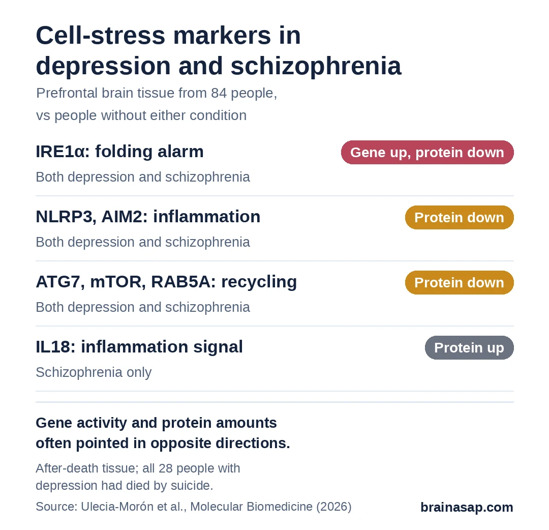
Results showed a mismatch. IRE1α gene activity was higher in both conditions, yet the IRE1α protein was lower.
Proteins for 2 inflammation sensors (NLRP3 and AIM2) and 3 cell-recycling helpers (ATG7, mTOR and RAB5A) were also lower in both groups.
Ulecia-Morón and colleagues suggested that IRE1α, which connects all 3 systems, might help explain why gene activity and protein amounts drifted apart.
Who Showed the Biggest Changes?
- Men with depression: most of the gene changes linked to the illness
- Women with schizophrenia: the widest range of gene changes
- Men overall: clearer drops in inflammation and recycling proteins
- Schizophrenia and suicide: higher activity of inflammation genes IL18 and CASPASE-5, plus more CASPASE-8 protein, in people who died by suicide
Why These Results Are Hard to Pin Down
- All depression cases were suicides: Some changes may reflect suicide or the hours before death rather than depression itself.
- After-death tissue only: Can't tell whether these changes came before the illness or followed it.
- Medicines varied: Antidepressants and antipsychotics in the blood weren't adjusted for in the analysis.
- Small groups: 28 per group, with fewer women, which weakens the comparisons between men and women.
- Limited protein tests: Only some markers could be measured as proteins because tissue was scarce.
Is IRE1α a Cause or a Consequence?
This study points to the brain's protein-folding alarm as a shared thread in depression and schizophrenia.
Testing it in people with depression who did not die by suicide, and following the gene and protein over time in animal or cell models, would show whether the mismatch helps drive these illnesses or is a result of them.
Citation: DOI: 10.1186/s43556-026-00514-4 Ulecia-Morón C, Bris ÁG, Inglada-Pérez L, Callado LF, Meana JJ, Martín-Hernández D, MacDowell KS, Figuero E, Caso JR, Leza JC. Transcription-protein dissociation reveals disrupted cellular stress pathways in the prefrontal cortex of depression and schizophrenia subjects. Molecular Biomedicine. 2026;7:138.
Study Design: Postmortem case-control study of dorsolateral prefrontal cortex (Brodmann area 9); RT-qPCR for inflammatory, autophagy and UPR genes; Western blot and ELISA for selected proteins; logistic and linear regression with sex, age and cause-of-death analyses.
Sample Size: 84 brains: 28 MDD (all suicide), 28 schizophrenia, 28 matched controls; Basque Institute of Legal Medicine, Bilbao, Spain.
Key Statistic: IRE1α mRNA OR 1.072 (95% CI 1.031–1.115) for MDD and 1.055 (1.024–1.087) for SCZ; IRE1α protein OR 0.917 (0.878–0.957) MDD, 0.707 (0.500–1.001, p = 0.05) SCZ. Lower protein in both disorders: NLRP3, AIM2, ATG7, mTOR, RAB5A.
Caveat: Observational postmortem design; all MDD subjects died by suicide; medication not included as covariate; modest and male-heavy sample; only a subset of markers measured as protein.