Optical coherence tomography (OCT) reveals significant retinal nerve fiber layer (RNFL) thinning in Parkinson’s disease (PD) patients, correlating with disease severity and suggesting OCT as a potential biomarker for PD diagnosis and monitoring.
Highlights:
- RNFL Thinning: Parkinson’s disease patients exhibit significant thinning of the retinal nerve fiber layer (RNFL) compared to non-PD individuals (p = 0.015).
- Disease Severity Correlation: The reduction in RNFL thickness correlates with the severity of Parkinson’s disease, as measured by the Unified Parkinson’s Disease Rating Scale (UPDRS III) (p = 0.04).
- Potential Biomarker: These findings support the use of OCT as a non-invasive tool for quantifying neurodegeneration and potentially serving as a biomarker for Parkinson’s disease.
- Clinical Implications: Assessing RNFL and ganglion cell complex (GCC) thickness via OCT could aid in the early diagnosis and ongoing monitoring of Parkinson’s disease progression.
Source: Frontiers in Neuroimaging (2024)
Major Findings: Retinal Thinning in Parkinsons Disease (2024)
1. Significant Thinning of the Retinal Nerve Fiber Layer (RNFL) in PD Patients
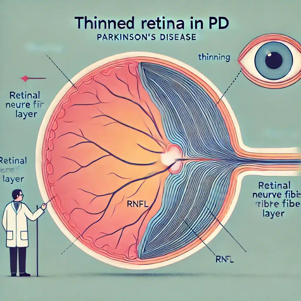
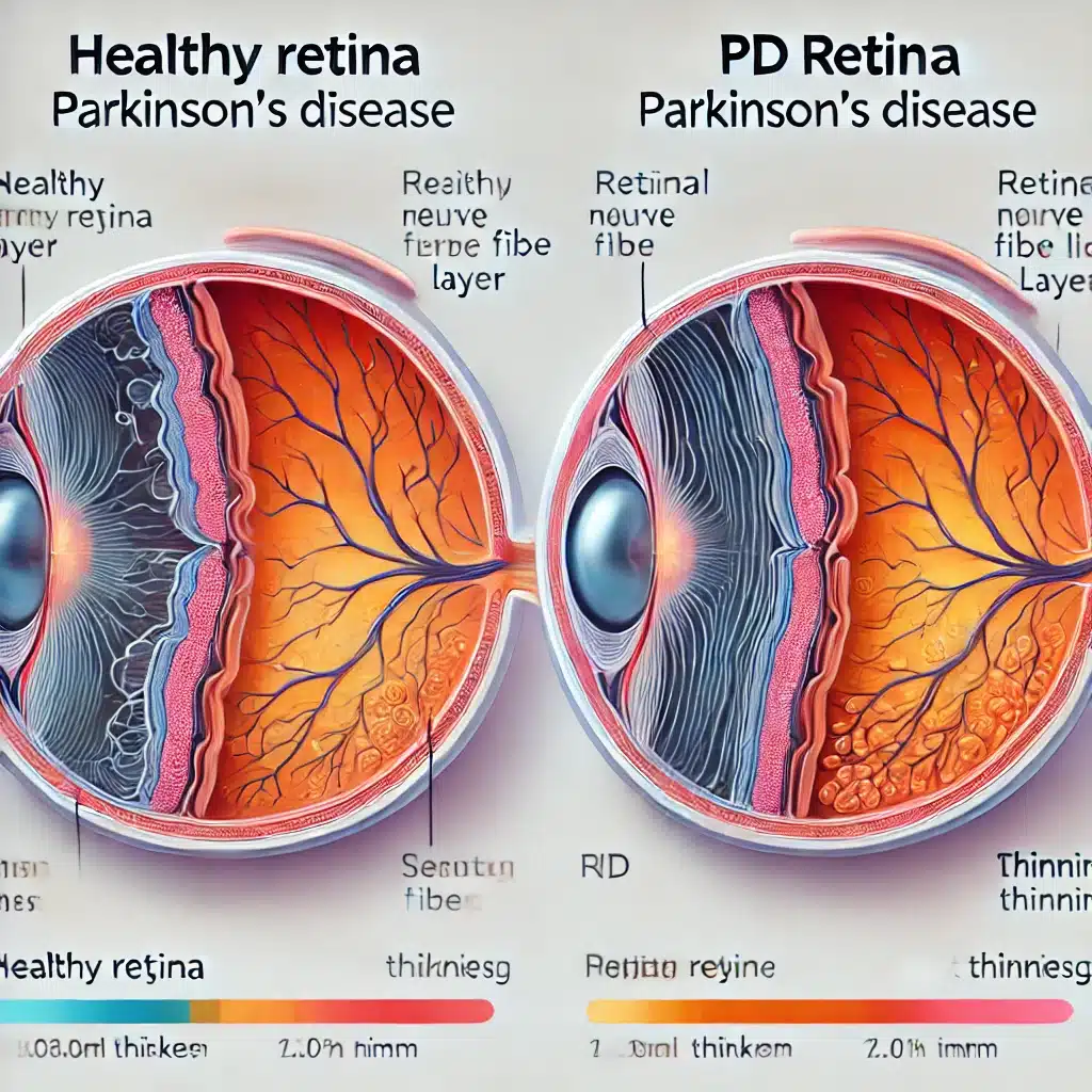
The study revealed that patients diagnosed with Parkinson’s disease (PD) show a significant reduction in the thickness of the retinal nerve fiber layer (RNFL) compared to individuals without PD.
This thinning was statistically significant (p = 0.015), suggesting that the neurodegenerative processes in PD extend beyond the brain to affect retinal structures.
The RNFL comprises the nerve fibers that transmit visual information from the eye to the brain, and its thinning reflects underlying neuronal loss associated with PD.
2. Correlation Between RNFL Thinning & Disease Severity
A critical finding of the study is the correlation between the degree of RNFL thinning and the severity of Parkinson’s disease.
The severity was assessed using the Unified Parkinson’s Disease Rating Scale (UPDRS III), which measures motor function impairment.
The study found a significant inverse correlation (p = 0.04), indicating that greater thinning of the RNFL is associated with more severe motor symptoms.
This suggests that RNFL thickness could serve as an indicator of disease progression in PD patients.
3. Optical Coherence Tomography (OCT) as a Potential Biomarker
The use of optical coherence tomography (OCT) in this study demonstrated its potential as a valuable tool for diagnosing and monitoring Parkinson’s disease.
OCT is a non-invasive imaging technique that provides high-resolution cross-sectional images of the retina.
The significant findings of RNFL thinning and its correlation with disease severity support the idea that OCT can be used to detect and quantify retinal neurodegeneration in PD patients.
This could lead to earlier diagnosis and better tracking of disease progression.
4. Matched Control Group Validates Findings
The study carefully matched PD patients with a control group of non-PD individuals based on age and sex.
This meticulous matching process ensured that the observed differences in RNFL thickness were specifically related to Parkinson’s disease rather than other confounding factors.
The control group confirmed that the RNFL thinning is a distinct characteristic of PD, reinforcing the reliability of the study’s findings.
5. Comprehensive Analysis & Robust Statistical Methods
The study utilized comprehensive statistical analyses to ensure the accuracy and reliability of its findings.
By employing methods such as the Shapiro–Wilk test for normal distribution and Pearson or Spearman correlation coefficients, the researchers validated the significance of their results.
Additionally, the use of multiple variable correction techniques, such as the Bonferroni adjustment, ensured that the observed correlations were not due to random chance but represented true associations between RNFL thinning and Parkinson’s disease severity.
(Related: Retinal Imaging Predicts Parkinson’s Up to 7 Years Before Diagnosis)
Study Overview: Retinal Changes in Parkinson’s Disease (2024)
The study examined retinal changes in Parkinson’s disease (PD) using optical coherence tomography (OCT) to assess the thickness of the retinal nerve fiber layer (RNFL) and ganglion cell complex (GCC).
The goal was to determine whether these retinal changes correlate with disease severity and could serve as biomarkers for early diagnosis and monitoring of PD.
Sample
The study included 30 patients diagnosed with Parkinson’s disease and 30 matched non-PD individuals.
Participants were matched based on demographic characteristics, and all underwent OCT and clinical evaluations to exclude other neurodegenerative and visual diseases.
Methods
- Design: Observational, retrospective study.
- Inclusion Criteria: Diagnosed with Parkinson’s disease, aged 45-85. MoCA score > 23. Intraocular pressure < 21 mmHg. No other neurodegenerative diseases, retinal/optic nerve conditions, ocular trauma, or significant refractive errors.
- Evaluations: Clinical assessment by a movement disorders specialist. Structured anamnesis and neurological examination including UPDRS III and Hoehn and Yahr classification. OCT measurements of the macula and optic nerve. Correlation analysis between clinical variables and OCT results using statistical methods (Shapiro–Wilk test, Pearson/Spearman correlation, Bonferroni adjustment).
Limitations
- Sample Size: The study was limited to 30 PD patients, which may not represent the broader PD population.
- Retrospective Design: Being retrospective, the study may have inherent biases related to patient selection and data recording.
- Disease Duration: The average disease duration of less than a decade may not capture the full spectrum of retinal changes in long-term PD.
- Medication Status: The majority of patients were evaluated in the ON medication state, which could influence UPDRS III scores and the interpretation of findings.
- Exclusion Criteria: Stringent exclusion criteria could limit the generalizability of results to all PD patients, especially those with comorbid ocular conditions.
Why Retinal Thinning Occurs in Parkinson’s Disease (Potential Reasons)

Retinal thinning in Parkinson’s disease (PD) is primarily attributed to the neurodegenerative processes that characterize the condition.
Dopaminergic Neuronal Loss
Parkinson’s disease involves the degeneration of dopaminergic neurons in the brain, particularly in the substantia nigra.
Dopamine is also present in the retina, where it plays a crucial role in visual processing.
In PD, the loss of dopaminergic neurons extends to the retina, leading to reduced dopamine levels and subsequent neuronal dysfunction and death.
Alpha-Synuclein Accumulation
PD is characterized by the abnormal accumulation of alpha-synuclein protein in the form of Lewy bodies within neurons.
This protein aggregation is not confined to the brain but also occurs in the retina.
The presence of alpha-synuclein in retinal cells disrupts normal cellular function, contributing to neuronal damage and retinal thinning.
Ganglion Cell Damage
Retinal ganglion cells, which form the optic nerve and transmit visual information to the brain, are particularly affected in PD.
The loss of these cells leads to thinning of the retinal nerve fiber layer (RNFL), which is composed of the axons of ganglion cells.
Damage to ganglion cells results in impaired visual processing and contributes to the overall thinning of the retina.
Inflammatory Processes
Inflammation is a known feature of Parkinson’s pathology.
Inflammatory responses in the retina can exacerbate neuronal damage and contribute to the thinning of retinal layers.
Chronic inflammation may lead to a hostile environment for retinal neurons, promoting their degeneration.
Oxidative Stress
Oxidative stress, resulting from an imbalance between the production of reactive oxygen species (ROS) and the body’s ability to detoxify them, is implicated in PD.
Retinal cells are highly susceptible to oxidative damage due to their high metabolic activity.
Increased oxidative stress can lead to cellular damage and death, contributing to retinal thinning.
Accuracy of Optical Coherence Tomography (OCT) in Diagnosing Parkinson’s Disease
Optical coherence tomography (OCT) has shown promising accuracy in diagnosing Parkinson’s disease (PD) by detecting retinal changes, specifically the thinning of the retinal nerve fiber layer (RNFL) and ganglion cell complex (GCC).
The study demonstrated that:
- Significant RNFL Thinning: PD patients exhibited a marked reduction in RNFL thickness compared to non-PD individuals (p = 0.015).
- Correlation with Disease Severity: The degree of RNFL thinning correlated with the severity of PD, as measured by the Unified Parkinson’s Disease Rating Scale (UPDRS III) (p = 0.04).
These findings indicate that OCT can reliably detect neurodegenerative changes associated with PD, making it a valuable diagnostic tool.
However, its accuracy depends on several factors, including the quality of the imaging equipment, the experience of the clinicians interpreting the results, and the specific characteristics of the patient population.
Early Diagnosis Potential
Early diagnosis of Parkinson’s disease is crucial for managing and potentially slowing the progression of the disease. OCT offers several advantages in this regard:
- Non-Invasive & Quick: OCT is a non-invasive imaging technique that provides rapid, high-resolution images of the retina, allowing for the early detection of retinal changes without discomfort to the patient.
- Detection Before Motor Symptoms: Retinal changes, such as RNFL thinning, may occur before the onset of motor symptoms. This means OCT could potentially identify PD at an earlier stage than traditional diagnostic methods, which typically rely on the appearance of motor symptoms.
- Monitoring Disease Progression: Regular OCT assessments can help track the progression of retinal changes over time, providing insights into the disease’s development and effectiveness of treatments.
Limitations in Early Diagnosis
While OCT shows promise for early diagnosis, there are limitations to consider:
- Overlap with Other Conditions: Retinal thinning can occur in other neurodegenerative and ocular conditions, potentially confounding the diagnosis of PD.
- Variability Among Patients: The extent of retinal changes may vary significantly among PD patients, and some individuals may not exhibit detectable retinal thinning in the early stages of the disease.
- Need for Complementary Tests: OCT should be used in conjunction with other diagnostic tools and clinical assessments to improve diagnostic accuracy and ensure comprehensive evaluation.
Conclusion: Retinal Thinning & Parkinson’s Diagnosis
The study demonstrated that optical coherence tomography (OCT) is an effective tool for detecting retinal changes in Parkinson’s disease (PD) patients, specifically showing significant thinning of the retinal nerve fiber layer (RNFL).
This retinal thinning correlates with the severity of the disease, suggesting that OCT could serve as a reliable biomarker for both early diagnosis and monitoring of PD progression.
The non-invasive nature and high-resolution imaging capabilities of OCT provide a valuable means of assessing neurodegeneration in PD, potentially allowing for earlier intervention.
Despite limitations such as sample size and variability in retinal changes among patients, these findings highlight the potential of OCT in enhancing the clinical management of Parkinson’s disease.
Further research with larger cohorts and longer follow-up periods is recommended to validate and expand upon these results.
References