
The Tiniest Neural Recorder: How Light-Powered Implants Can Monitor the Brain for a Year
TL;DR: A nanometer-scale wireless implant (MOTE) powered by light recorded stable brain activity in mice for a full year without tethers or degradation.
Recording brain activity for days is routine. Recording for weeks is impressive. Recording for an entire year from a wireless implant smaller than a grain of salt seemed impossible until now.
A team of biomedical engineers has announced a breakthrough: the MOTE (Microscale Optoelectronic Tetherless Electrode), a subnanolitre neural implant powered entirely by light. It recorded stable brain activity in awake mice for 365 days straight — no wires, no batteries, no signal degradation.
The device eliminates the mechanical tether that causes scarring and drift, and uses a dual-wavelength optical system to power the implant and stream neural data wirelessly. It represents a leap toward understanding how the brain works over months and years, and how neurological and psychiatric disorders evolve in real time.
Key Findings
- Subnanolitre per channel: MOTE measures <0.001 nanolitres per recording channel—orders of magnitude smaller than RF or ultrasound-based tetherless implants (which typically displace microlitres or greater)—minimizing tissue damage and foreign-body response.
- Photovoltaic light-emitting diode (PVLED): The device uses a single PVLED operating in time-division multiplexing: it harvests power as a PV cell 93.4% of the time and emits data-encoded infrared pulses for 0.06% of the time, eliminating traditional power/communication crosstalk.
- Safe irradiance: Incident LED irradiance is limited to <70 mW/mm², well below the threshold of 250 mW/mm² for thermal brain damage, enabling safe chronic illumination through intact skull.
- Pulse-position modulation (PPM): On-chip PPM encoding compresses neural signals into bright, short pulses, achieving low noise (14.8 µVrms) and high temporal resolution (>10 kHz bandwidth) with minimal power consumption (1 µW per channel).
- 365 days of stable recording: MOTE successfully recorded local field potentials (LFPs) and action potentials from barrel cortex neurons in awake mice for a full year, showing stable signal quality and excellent tracking of sensory evoked responses.
- In vitro validation: Using iPSC-derived cardiomyocytes confirmed faithful recording of electrophysiological activity, with PPM decoding accurately reproducing voltage dynamics including pharmacological modulation.
Source: Nature Electronics (2025) | Lee, Molnar, et al.
The Tether Problem: Why Wireless Matters for Chronic Recording
Neuroscientists have long relied on wired neural implants—electrodes connected by hair-thin tethers to external amplifiers and computers. Wires work, but they inflict cascading costs:
- Mechanical friction: Every micro-movement of the head, muscle twitch, and breath creates friction between the rigid tether and surrounding brain tissue
- Tissue damage: Over weeks, this causes scarring, glial activation, and electrode drift
- Acute success, chronic failure: Recording for minutes to hours is reliable; stable recording for months remains unsolved
Wireless implants promise freedom from the tether, but size and power become the bottleneck. Existing wireless approaches have two main limitations:
- Radiofrequency (RF) systems — limited by wavelength physics. The smaller the implant, the harder it is to couple RF energy efficiently. Most RF devices displace microlitres of brain tissue per channel, causing significant damage.
- Ultrasound-powered systems — face similar size constraints and tissue displacement issues. Most aren’t fully embedded; they sit atop the skull on a pedestal.
MOTE takes a different approach entirely: light. Optical wavelengths enable a nanolitre-scale implant with key advantages:
- Short wavelengths: 623 nm for the power-delivering red laser; 825 nm for data-emitting infrared
- Coupling efficiency: Short wavelengths allow subnanolitre scale without sacrificing energy transfer
- Tissue tolerance: The brain tolerates light at these wavelengths with minimal scatter
- Safety margins: Power densities stay well below thermal damage thresholds
- Result: An implant so small it causes virtually no tissue disruption
Clever Optoelectronics: Powering and Communicating Simultaneously
The engineering elegance of MOTE lies in one component doing two jobs. At its core is an AlGaAs photovoltaic light-emitting diode (PVLED) that operates in a clever dual role:
- Power harvesting: Acts as a photovoltaic cell, harvesting power from a red laser (623 nm)
- Data transmission: Emits data as infrared light (825 nm) when driven by on-chip circuitry
- Time-division multiplexing: The diode harvests power 93.4% of the time and emits data pulses 0.06% of the time, preventing crosstalk
- Dual-wavelength isolation: Red powers in, infrared sends data out, avoiding crosstalk problems
How It Encodes Brain Signals
Rather than varying light brightness (which is noisy), MOTE uses pulse-position modulation (PPM) — encoding information in the timing of brief, bright pulses rather than their intensity. A reference pulse fires at a fixed interval; an encoding pulse follows at a delay proportional to neural voltage. The time gap is the message. It’s the same encoding scheme satellites use: extraordinarily noise-resistant.
The numbers are remarkable. Noise floor: 14.8 µVrms. Bandwidth: over 10 kHz. Power consumption: 1 microwatt per channel — a single AAA battery could theoretically power 2,700 MOTEs for a year. The external laser stays below 70 mW/mm², well under the 250 mW/mm² threshold for thermal brain damage.
Building the Impossible: Fabrication at Nanoscale
Constructing a subnanolitre optoelectronic implant means bonding two fundamentally different materials — silicon CMOS circuitry and gallium arsenide optoelectronics — with mismatched crystal structures. Three fabrication innovations make it work:
- Vacuum annealing at 300°C removes organic residues from bonding surfaces, creating clean silicon-semiconductor interfaces without thermal damage.
- Atomic layer deposition (ALD) wraps the device in ultrathin protective layers (SiO2, Si3N4, Al2O3) under 1.5 micrometres, shielding it from the biological environment.
- Platinum light shielding — a 94 nm platinum layer covers circuitry while exposing optical components, acting like a Faraday cage to block stray photons and noise.
Nearly 100 MOTEs are fabricated in parallel on a single wafer, scalable to thousands per square centimetre. Each is handled using a pulled glass micropipette and sterilized before implantation.
Proof of Concept: One Year of Stable Recording in Awake Mice
The first validation came in vitro. Cultured iPSC-derived cardiomyocytes were plated on MOTEs in a Petri dish. The results demonstrated three key proofs:
- Baseline recording: The MOTE faithfully recorded cell electrical activity
- Dynamic response: When researchers applied isoproterenol (to speed beating) or blebbistatin (to slow it), MOTE output tracked changes in real time
- Signal fidelity: PPM decoding accurately reconstructed voltage waveforms, proving optical encoding worked without information loss
The critical test came in vivo. Six mice were surgically implanted with MOTEs in the barrel cortex, the sensory region processing whisker touch. The experimental design included:
- Cortical implants: Four mice had MOTEs in cortical layers
- Surface implants: Two mice had implants on the brain surface for electrocorticographic (ECoG) signals
- Sensory protocol: Whisker stimulation with a motorized rod during awake, head-fixed recordings
- Duration: Neural responses recorded for 365 days
MOTE recorded two signal types:
- Local field potentials (LFPs): slow oscillations from population-level neural activity
- Action potentials: rapid voltage spikes from individual neurons
Signal stability exceeded expectations across three dimensions:
- Duration: Stable for the full year
- Evoked response clarity: Whisker stimulation responses were clear on day 4, day 123, and day 161—genuine neural recording
- Tissue biocompatibility: Minimal scarring around implant sites, confirming the device’s subnanolitre footprint and biocompatible encapsulation
What This Unlocks: Long-Term Neural Dynamics and Disease Models
Year-long recording opens previously inaccessible doors. MOTE enables investigation of slow neurological processes:
- Neurodevelopmental disorders: Track how neural circuits fail to mature
- Cognitive decline: Watch how memory systems degrade over months
- Psychiatric disease: Observe how mood-regulating networks destabilize
Neuroscientists previously inferred neural basis from snapshots. MOTE lets them watch the brain over timescales matching symptom development.
Research Applications
- Disease progression — Chronic epilepsy, Alzheimer’s neuroinflammation, and other slow disorders now become trackable without tether-induced damage.
- Scaling down — With miniaturization, MOTEs could record entire small-organism nervous systems (C. elegans: 302 neurons) non-invasively.
- Scaling up — Hundreds of MOTEs across brain regions create wireless brain maps during complex behaviors like social interaction or decision-making.
Clinical Potential
Clinical translation is farther off but tantalizing:
- Brain-computer interfaces: Wirelessly powered through intact scalp to restore communication for paralyzed patients
- Seizure monitoring: Chronically implanted MOTEs detect seizure precursors, triggering closed-loop neuromodulation before seizures begin
- Distributed recording: Subnanolitre size means many MOTEs coexist without tissue disruption, mapping networks rather than single focal areas
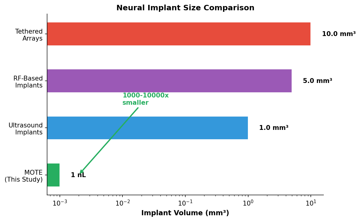
Image file: mote-neural-implant.png — MOTE device schematic showing dual-wavelength optical architecture (623 nm power, 825 nm data), on-chip PPM encoder, and comparative size with human hair and other neural recording technologies.
A New Era in Neuroscience
The ability to record brain activity chronically, wirelessly, and at subnanolitre scale is a watershed moment. Tethered recording remains important for acute experiments but can no longer be the standard for chronic work. MOTE shifts the paradigm. The smallest implant has opened the biggest window into the working brain.
Citation: Lee, S., Molnar, A.C., et al. (2025). A subnanolitre tetherless optoelectronic microsystem for chronic neural recording in awake mice. Nature Electronics, 8, 1259–1271. DOI: 10.1038/s41928-025-01484-1
Key innovations: Photovoltaic light-emitting diode (PVLED) for simultaneous power and communication; pulse-position modulation (PPM) encoding; atomic layer deposition (ALD) encapsulation; time-division multiplexing; 365-day chronic in vivo validation in awake behaving mice.
Affiliation lead: Alyosha C. Molnar, Cornell University; Sunwoo Lee (corresponding), Nanyang Technological University, Singapore.