TL;DR: A 2026 meta-analysis in Psychological Medicine found postpartum depression (PPD) brain-activity differences across default-mode, limbic, and sensorimotor regions, with spatial overlap in serotonin, dopamine, and vesicular acetylcholine transporter maps.
Key Findings
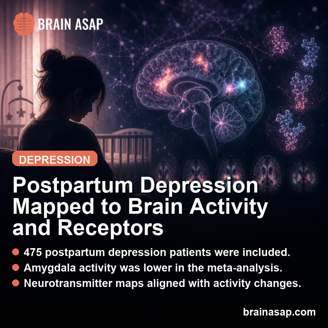
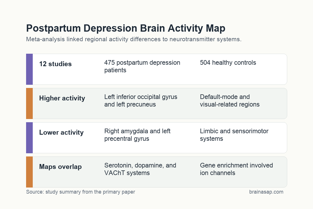
- 12 imaging studies pooled: The meta-analysis included 475 postpartum depression patients and 504 healthy controls.
- Higher activity appeared in two regions: PPD was linked to increased resting-state functional activity in the left inferior occipital gyrus and left precuneus.
- Lower activity appeared in two regions: PPD was linked to decreased resting-state functional activity in the right amygdala and left precentral gyrus.
- Three brain systems were implicated: The regions mapped onto default-mode network, limbic, and primary sensorimotor systems.
- Neurotransmitter maps overlapped: The spatial pattern overlapped with serotonergic, dopaminergic, and vesicular acetylcholine transporter (VAChT) systems.
- Gene enrichment involved ion channels: Allen Human Brain Atlas analysis linked PPD-related regions to transmembrane transport, gated/passive channels, and channel complexes.
Source: Psychological Medicine (2026) | Chen et al.
Postpartum depression (PPD) is a depressive disorder that occurs after childbirth and can affect mood, bonding, sleep, cognition, and safety. Resting-state brain imaging studies have tried to identify its neural pattern, but individual studies have not always agreed.
This meta-analysis pooled resting-state functional imaging findings and then asked whether the resulting brain map lined up with known neurotransmitter and gene-expression maps.
12 Resting-State Functional MRI Studies Covered 979 Participants
The researchers reviewed resting-state functional imaging studies comparing postpartum depression patients with healthy controls. The final meta-analysis included 12 studies, with 475 PPD patients and 504 controls.
- PPD group: 475 postpartum depression patients contributed imaging data across the pooled studies.
- Control group: 504 healthy controls provided the comparison baseline.
- Method: Resting-state functional MRI compared spontaneous activity while participants were not performing a task.
Resting-state functional MRI (fMRI) measures spontaneous brain activity when a participant is not performing a task. It can identify regions or networks whose baseline activity differs between groups.
The analysis used SDM-PSI, a voxel-based meta-analytic method. In plain terms, it combined reported imaging coordinates across studies to identify brain regions where PPD differences appeared consistently.
This pooling step is valuable because individual PPD imaging studies are often small. A meta-analysis cannot remove every difference among studies, but it can ask whether a regional pattern keeps appearing across independent samples.
The researchers then added neurotransmitter and gene-expression mapping, which moves the result from a location list toward a mechanistic hypothesis.
PPD Showed Higher Activity in Precuneus and Inferior Occipital Gyrus
Two regions showed increased resting-state functional activity in postpartum depression: the left inferior occipital gyrus and the left precuneus.
The precuneus is part of the default mode network, a system involved in internally directed thought, self-referential processing, and memory-related mental activity. The inferior occipital gyrus is involved in visual processing.
Higher activity in these regions should not be read as “more brain function” in a simple good-or-bad sense. It means the baseline activity pattern differed from controls in regions connected to perception and internally oriented processing.
The precuneus finding is especially relevant because postpartum depression often includes rumination and altered self-focused thinking. The meta-analysis cannot assign that symptom to the precuneus, but it places the region in the set of systems that deserve closer longitudinal testing.
The occipital finding is less clinically obvious, yet visual regions can participate in broader networks involved in attention, salience, and internally generated imagery.
Right Amygdala and Left Precentral Gyrus Activity Was Lower
Two regions showed lower resting-state functional activity: the right amygdala and the left precentral gyrus.
The amygdala is central to emotion salience, threat processing, and affective learning. The precentral gyrus contains primary motor cortex and is part of the sensorimotor system.
Together, the higher and lower regions suggest PPD is not a single-location disorder. The pattern spanned emotional, internally directed, visual, and sensorimotor systems.
The distribution fits the clinical breadth of postpartum depression. Mood symptoms, anxiety, threat sensitivity, disrupted sleep, slowed or agitated behavior, and altered self-focused thinking can all occur in the postpartum period.
The imaging pattern does not assign one symptom to one region. It does show that pooled PPD differences touched systems plausibly related to emotion, self-processing, and action readiness.
Serotonin, Dopamine, and VAChT Maps Overlapped With PPD Regions
The second layer of the study used JuSpace, a toolbox that compares brain findings with nuclear-imaging-derived neurotransmitter maps.
The PPD activity pattern spatially overlapped with serotonergic and dopaminergic systems. It also overlapped with VAChT, the vesicular acetylcholine transporter, a marker related to acetylcholine signaling.
This does not prove that serotonin, dopamine, or acetylcholine changes cause postpartum depression. It does suggest the affected regions sit in parts of the brain where those neurotransmitter systems are relevant.
The neurotransmitter overlap is relevant because postpartum depression treatment already intersects with monoamine systems through antidepressants, while acetylcholine and sensorimotor findings may open additional mechanistic questions.
Gene Enrichment Involved Ion Channel Function
The researchers then used the Allen Human Brain Atlas to compare the imaging map with regional gene-expression patterns.
PPD-related gene enrichment involved transmembrane transport, gated and passive channels, and channel complexes. Those terms describe ion-channel function, the machinery that helps neurons regulate electrical activity and signaling.
The gene-expression result should be read as hypothesis-generating. It links a spatial imaging pattern to broad molecular categories, but it does not identify a diagnostic gene test or treatment target by itself.
Ion-channel enrichment is relevant because channels shape neuronal excitability. If future work replicates this link, researchers could test whether altered excitability helps explain the resting-state activity differences.
For now, the gene layer mainly helps prioritize biology for follow-up rather than settle the mechanism.
Meta-Analysis Helps but Does Not Solve PPD Biomarkers
The study improves on single small imaging studies by pooling evidence. Still, the total sample is under 1,000 participants, and resting-state imaging studies can differ in scanner protocols, preprocessing, symptom measures, medication status, postpartum timing, and sleep disruption.
PPD is also clinically heterogeneous. Hormonal changes, prior depression history, social stress, sleep deprivation, breastfeeding, obstetric complications, and anxiety symptoms can all shape postpartum mood and brain function.
Medication status is another practical issue. Antidepressant exposure, sleep medication, or untreated symptom severity could affect resting-state activity, and small studies may not be able to model those factors cleanly.
Postpartum timing also matters because the biology of 6 weeks after delivery may differ from the biology of 9 months after delivery.
Because of those limits, the findings are better treated as a mechanistic map than as a clinical diagnostic tool.
A More Specific Brain-System Hypothesis
The strongest contribution is specificity. Instead of saying postpartum depression affects “the brain,” the meta-analysis identifies a pattern involving default-mode, limbic, visual, and sensorimotor regions.
By linking those regions to neurotransmitter maps and ion-channel gene-expression categories, the study gives researchers a more testable set of hypotheses for future PPD work.
The next step is larger longitudinal imaging that tracks symptom change, treatment response, sleep, hormones, and postpartum timing in the same participants.
That design would clarify whether the mapped regions change with recovery, treatment response, or postpartum timing rather than only distinguishing pooled patient and control groups.
It would also help separate illness biology from sleep loss, medication exposure, and postpartum stage.
Citation: DOI: 10.1017/S0033291726103651; Chen et al., intrinsic regional brain activity differences and neurotransmitter-gene mapping in postpartum depression, Psychological Medicine 2026.
Study Design: Whole-brain voxel-wise meta-analysis of resting-state functional imaging studies with neurotransmitter-map and gene-expression analyses.
Sample Size: 12 studies; 475 postpartum depression patients and 504 healthy controls.
Key Result: PPD showed increased activity in left inferior occipital gyrus and left precuneus, decreased activity in right amygdala and left precentral gyrus, and overlap with serotonergic, dopaminergic, and VAChT systems.
Caveat: The pooled imaging evidence is mechanistic and hypothesis-generating, not a diagnostic test for individual postpartum patients.