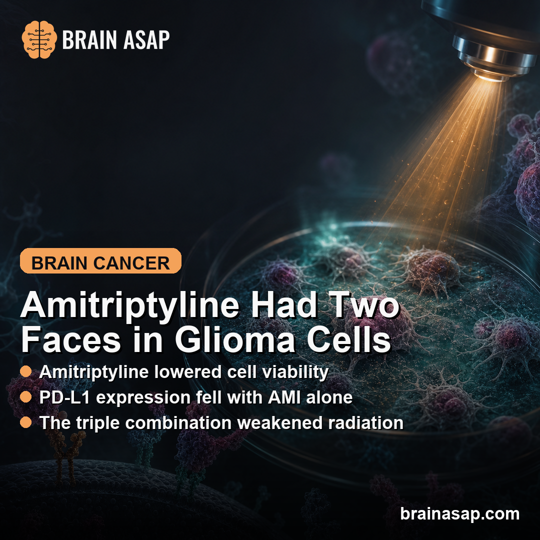
TL;DR: A 2026 in vitro study in Molecular and Clinical Oncology found that amitriptyline reduced C6 glioma cell viability and PD-L1 checkpoint expression, but the same drug weakened the cell-killing effect of radiation when combined with temozolomide.
Key Findings
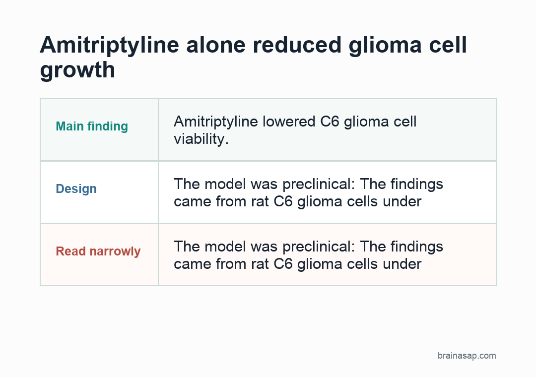
- Amitriptyline alone reduced glioma cell growth: In non-radiated C6 glioma cultures, 10 micromolar amitriptyline lowered viability by about 36% and increased cell mortality by about 23%.
- Radiation was stronger by itself: A single 10 Gy radiation exposure lowered viability by about 67% and increased mortality by about 57% versus untreated non-radiated cells.
- PD-L1 moved in opposite directions: Amitriptyline lowered PD-L1 expression, while temozolomide and radiation increased it.
- The triple combination raised concern: In radiated cultures, amitriptyline plus temozolomide reversed much of the radiation effect and restored cell viability toward untreated levels.
- The model was preclinical: The findings came from rat C6 glioma cells under hypoxic culture conditions, not from patients with glioma.
Source: Molecular and Clinical Oncology (2026) | Bielecka-Wajdman et al.
Amitriptyline Was Tested as a Glioma Drug-Repurposing Candidate
Amitriptyline is a tricyclic antidepressant also used for chronic and neuropathic pain.
In glioma care, that overlap is clinically relevant because patients may receive antidepressants or pain treatments alongside surgery, radiation, and chemotherapy.
The study asked a narrow preclinical question: what happens when C6 rat glioma cells are exposed to amitriptyline alone or together with temozolomide, the standard chemotherapy drug often used in glioma treatment, and 10 Gy radiation?
Researchers cultured the cells under low-oxygen conditions meant to resemble part of the tumor environment. They then measured viability, mortality, proliferation, colony formation, and PD-L1, a checkpoint protein that can help tumor cells avoid immune attack.
The result had two sides. Amitriptyline looked harmful to glioma cells when used alone.
The combination with radiation and temozolomide looked less favorable, because the paired drugs weakened the radiation effect in this cell model.
The study does not show that patients with glioma should start or stop amitriptyline. It shows why drug interactions around cancer treatment need direct testing rather than assumptions based on a single drug’s effects.
C6 Glioma Cells Were Exposed to Drug and Radiation Combinations
The experiment used C6 rat glioma cells, a common laboratory model for glioma biology. Cells were treated for 72 hours with amitriptyline, temozolomide, both drugs, radiation, or drug-radiation combinations.
The main treatment conditions were:
- Amitriptyline: 10 micromolar, a concentration the study authors described as relevant to brain exposure during treatment.
- Temozolomide: 1 millimolar, used as the chemotherapy comparator and combination partner.
- Radiation: A single 10 Gy exposure, followed by the same 72-hour observation period.
- Combination groups: Drug-treated cultures were compared with radiated and non-radiated controls.
Several readouts were included because no single assay captures the whole cell response.
MTT measured mitochondrial activity as a viability readout, Trypan blue counted dead cells, BrdU measured proliferation, microscopy assessed colonies, and an enzyme assay measured PD-L1 expression.
Triplicate measurements were used for the functional tests, and figure data were reported as mean plus or minus SEM from three independent experiments, with n=9 per group.
Amitriptyline Alone Lowered Viability and PD-L1 Expression
In non-radiated glioma cultures, amitriptyline had a clear anticancer-direction result. Cell viability fell by about 36% compared with untreated non-radiated cultures, while cell mortality rose by about 23%.
Proliferation also moved in the same direction. BrdU-positive cells fell by about 26% with amitriptyline, while temozolomide and the amitriptyline-plus-temozolomide condition each reduced proliferation by about 34%.
The checkpoint result was different from the temozolomide result:
- Amitriptyline lowered PD-L1: Non-radiated amitriptyline-treated cells showed about a 12% decrease in PD-L1 expression.
- Temozolomide increased PD-L1: Temozolomide raised PD-L1 by about 11% in non-radiated cells.
- Radiation increased PD-L1: Radiation alone raised PD-L1 by about 11% compared with non-radiated controls.
The PD-L1 pattern is clinically relevant but still early. PD-L1 is involved in immune checkpoint signaling.
This experiment measured direct effects in cultured glioma cells. It did not test whether amitriptyline improves immune response inside a living tumor.
Amitriptyline Plus Temozolomide Weakened Radiation’s Cell-Killing Effect
Radiation alone produced the strongest cell-killing result. A single 10 Gy radiation exposure lowered viability by about 67%, increased mortality by about 57%, and reduced proliferation by about 60% compared with untreated non-radiated cells.
The interaction changed when drugs were added to radiated cultures. Amitriptyline or temozolomide alone attenuated the radiation effect, and the amitriptyline plus temozolomide condition produced the most concerning reversal.
In radiated cultures treated with both drugs, cell viability became comparable to untreated non-radiated cells. Mortality also moved back toward the non-radiated control pattern, and proliferation increased by about 35% compared with non-radiated cultures treated with the same drug pair.
Microscopy matched the assay results. Radiation alone left fewer live cells after 72 hours. The radiated cultures treated with temozolomide or amitriptyline plus temozolomide showed fuller confluence and larger colony formation.
Amitriptyline is not categorically harmful in glioma based on this experiment. The result shows that a drug can look anticancer by itself and still interfere with a separate treatment combination under specific laboratory conditions.
The PD-L1 Result Needs In Vivo Testing Before Clinical Claims
PD-L1 is a programmed death-ligand checkpoint protein; tumor PD-L1 can bind immune-cell PD-1 and reduce antitumor immune activity.
Lower PD-L1 expression after amitriptyline would be more important if it also occurs in living tumors with immune cells, microglia, blood vessels, and treatment metabolites.
This study did not test that environment.
Several limits keep the finding preclinical:
- Cell-line model: C6 rat glioma cells do not reproduce the full biology of human glioblastoma.
- No immune microenvironment: The culture system cannot show how microglia, T cells, cytokines, or tumor niches respond.
- Single radiation design: One 10 Gy exposure is not the same as a full clinical radiotherapy course.
- No active metabolites: Cultured cells were not exposed to the full metabolism that occurs in patients.
Those limitations define the result.
The study supports more testing of amitriptyline’s glioma-cell and checkpoint effects, but it also warns that timing and combination design may determine whether the effect helps or hurts.
The next useful step would be an in vivo glioma model that tests amitriptyline timing separately from temozolomide and radiation timing.
That model would show whether the PD-L1 decrease survives in a tumor environment and whether the radiation-interference pattern appears outside the culture dish.
Citation: DOI: 10.3892/mco.2026.2947. Bielecka-Wajdman et al. Two faces of Amitriptyline in an in vitro study on C6 glioma cells: The effects of Amitriptyline and its combination with Temozolomide and radiation. Molecular and Clinical Oncology. 2026;24:38.
Study Design: In vitro C6 rat glioma cell experiment testing amitriptyline, temozolomide, radiation, and their combinations.
Sample/Model: C6 rat glioma cells cultured under hypoxic conditions, with functional assays reported from three independent experiments and n=9 per group.
Key Statistic: Amitriptyline alone lowered non-radiated cell viability by about 36%, while amitriptyline plus temozolomide reversed much of the 10 Gy radiation effect in radiated cultures.
Caveat: The experiment used a cell-line model, so patient-level safety or treatment-sequencing conclusions require animal and clinical evidence.