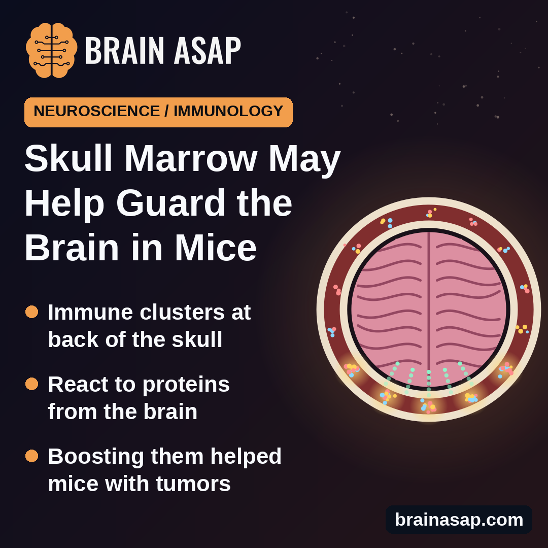
TL;DR: In mice, the bone marrow inside the skull holds small immune hubs that pick up proteins leaking out of the brain and react to them, including proteins from brain tumors. Boosting those hubs with a gel placed under the scalp helped mice with brain tumors live longer.
Key Findings
- Lymph-node-like clusters in the skull: T cells, B cells and antigen-presenting cells grouped together in skull marrow, mostly at the back of the head.
- More active than other bones: Skull marrow had more of these cells than marrow from the breastbone or thighbone.
- Brain proteins reached them: A marker protein made by brain cells turned up in skull marrow and switched on matching immune cells there.
- Blocking hurt, boosting helped: In mice with brain tumors, a scalp gel that blocked the hubs shortened survival; a three-part gel that boosted them lengthened it.
- One drug alone backfired: The main drug by itself did not help and tended to shorten survival.
Source: Nature (2026) | Park et al.
For a long time the brain was described as walled off from the immune system. That picture has been falling apart, and one of the more surprising pieces is the skull itself. Tiny channels connect the bone marrow in the skull to the fluid around the brain.
A team at Washington University in St. Louis asked what that skull marrow is actually doing. In mice, the researchers found it contains small immune training grounds that keep watch on the brain, and that these hubs matter in brain cancer.
The Skull’s Marrow Looks Like a Small Lymph Node
Bone marrow is mostly known as a factory for new blood and immune cells. Lymph nodes are where those cells learn to recognize a specific threat. In lymph nodes, helper T cells meet B cells in spots called germinal centers, where B cells sharpen their antibodies.
Using single-cell sequencing, flow cytometry and imaging of cleared skull bone, the researchers found the same ingredients in mouse skull marrow:
- Helper T cells of the type that coach B cells, at a higher share than in breastbone or thighbone marrow
- Germinal-center-like B cells and antibody-making cells, also more common than in other marrow
- Clusters of T and B cells with the support cells and signals of a germinal center
- Signs of antibody refinement, including expanded clones and mutated antibody genes
The clusters sat mainly in the back of the skull, in the occipital and interparietal bones. They were already present in mice 7 days after birth. A public dataset of human skull marrow also contained similar helper T cells, though in low numbers.
Proteins From the Brain Reached the Skull Hubs
To test what these hubs respond to, the team made mouse neurons produce a harmless marker protein. A month later, the protein had drained to the skull marrow, strongest at the back of the skull, and to the membranes around the brain. It was not detected in the blood.
Immune cells built to recognize that protein were then injected into the mice. One week later, these cells became active and formed germinal-center-like cells in the skull marrow. Comparable cells in the breastbone marrow, spleen and neck lymph nodes showed no significant change.
Mice that lacked a water channel needed for the brain’s fluid-clearing (glymphatic) system delivered less of the protein to the skull and had weaker responses there. That points to normal fluid drainage as the route.
Brain Tumors Switched On the Same Hubs
The researchers repeated the test with a mouse brain tumor (glioma) engineered to carry the marker protein. Immune cells that recognize it multiplied and activated in the skull marrow, mostly at the back. When the same tumor grew under the skin of the flank, the response appeared in nearby lymph nodes, not the skull.
The skull response was fast. Two days after the cells were injected, activated T cells showed up only in skull marrow. It also did not depend on the usual route: tying off the neck lymph nodes did not reduce it, and it still appeared in mice without lymph nodes or a spleen, though weaker.
A Scalp Gel Changed How Long Mice Survived
Next, the team placed a slow-release gel under the scalp to act mainly on the skull marrow below it.
- Blocking the hubs: A gel with an antibody that blocks a key T cell-B cell signal (CD40L) shortened survival in tumor-bearing mice, 10 per group (P = 0.0065). Fewer immune cells reached the tumor.
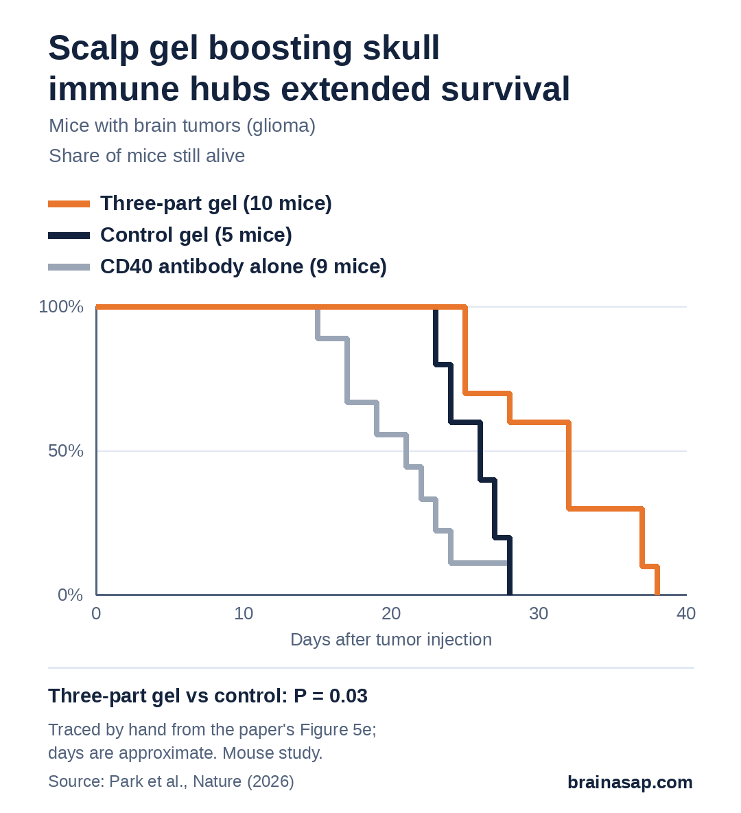
- Boosting the hubs: A gel with three immune boosters (an antibody that activates CD40 plus the signaling proteins IL-21 and interferon gamma) lengthened survival compared with control gel (P = 0.03).
- One booster alone: The CD40-activating antibody by itself did not help (P = 0.10 vs control), and these mice tended to die earliest. The authors suspect it favored B cells that dampen immunity.
The three-part gel did not work in mice that lacked B cells or antibody-making cells, which suggests antibodies are doing much of the work. In treated mice, killer T cells and natural killer cells inside the tumor became more active.
What a Mouse Study Can’t Say About People
- Mice only: Human skull marrow showed similar helper T cells, but in low numbers; whether the hubs work the same in people is untested.
- Small survival groups: 5 to 10 mice per group; treated mice lived days longer, not cured.
- Engineered tumors and proteins: Marker proteins made the responses easy to track but are not natural brain proteins.
- Not the only route: Authors note lymph nodes and spleen still add to the response; mice lacking them did worse.
- Single drugs can backfire: CD40 activation alone tended to shorten survival.
Next Step: Finding These Hubs in Human Skulls
The study adds the skull to the list of places where the immune system keeps an eye on the brain, and it suggests a local target that could be reached without going through the skull. Nothing here is a treatment for people with brain tumors.
The open question is whether human skull marrow, especially at the back of the head, holds the same working hubs, and whether they respond to real tumors in patients.
Citation: DOI: 10.1038/s41586-026-10951-4. Park JH, Abramishvili D, Davanzo GG, et al. Functional role of skull lymphoid structures in CNS immunosurveillance. Nature. 2026.
Study Design: Laboratory study in mice: single-cell sequencing, flow cytometry, imaging, antigen tracking and brain tumor (glioma) survival experiments, plus reanalysis of public human skull marrow data.
Sample Size: Mostly 3 to 10 mice per experiment; survival groups of 10 vs 10 (blocking) and 5, 9 and 10 (boosting).
Key Statistic: Blocking skull hubs shortened survival (P = 0.0065); three-part scalp gel lengthened it vs control (P = 0.03).
Caveat: Mouse study with engineered tumors and marker proteins; human relevance not yet tested.