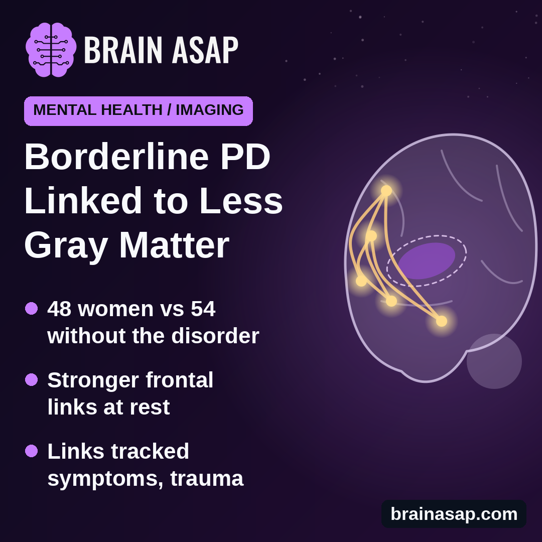
TL;DR: Brain scans of 48 women with borderline personality disorder showed less gray matter in the insula and parts of the frontal lobe than in 54 women without it. Some of those frontal areas were more strongly in sync with other brain regions, and that syncing was linked to symptom severity, self-harm and childhood trauma.
Key Findings
- Less gray matter: In the insula on both sides and in the middle and upper frontal lobe.
- Stronger resting links: From right frontal areas to left areas behind the eyes and at the front of the temporal lobe.
- Linked to symptoms: Stronger links went with more severe borderline symptoms and more self-harm.
- Childhood maltreatment linked too: Different types of abuse and neglect linked to different connection patterns.
- Gray matter itself not linked to symptoms or trauma scores.
Source: European Archives of Psychiatry and Clinical Neuroscience (2026) | Flasbeck et al.
Borderline personality disorder brings intense, fast-changing emotions, unstable relationships and, for many, self-harm. Childhood abuse and neglect are common in people with the diagnosis. Brain imaging studies have found differences in the frontal lobe and the insula, but rarely asked how brain structure and brain activity fit together.
Researchers at Ruhr University Bochum in Germany did both in the same group of women.
Two Scans, Same Women
The study included women receiving inpatient treatment for borderline personality disorder and women with no mental health history recruited online. Everyone had two kinds of MRI:
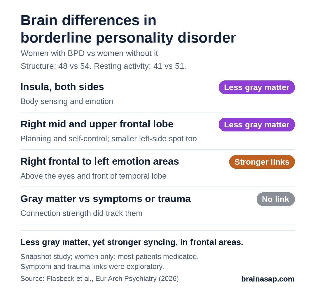
- Structure scan: Measured gray matter volume across the whole brain. 48 patients and 54 comparison women were analyzed.
- Resting scan: About 5 minutes of lying still, eyes open, to see which brain areas rise and fall in activity together. 41 patients and 51 comparison women were analyzed.
The areas that differed in structure became starting points for the activity analysis. Participants also filled in questionnaires on borderline symptoms, self-harm and childhood maltreatment.
Less Gray Matter in the Insula and Frontal Lobe
Patients had less gray matter than comparison women in:
- Insula, both sides: Deep area involved in sensing the body’s inner state and in emotion.
- Middle and upper frontal lobe: Areas involved in planning and self-control, mostly on the right.
- Two small spots: A patch at the back of the brain and one under the frontal lobe.
No area had more gray matter in patients. Gray matter volume was not linked to symptom, self-harm or trauma scores.
Frontal Areas Were More in Sync
The right frontal areas with less gray matter were, at rest, more strongly in sync with several areas on the left side: the orbitofrontal cortex just above the eyes, the front tip of the temporal lobe, and other frontal regions. These areas help with judging rewards, emotions and social situations.
Links With Symptoms, Self-Harm and Childhood Trauma
In exploratory analyses, stronger connections between the right frontal areas and the orbitofrontal cortex went with more severe borderline symptoms. Higher self-harm scores went with stronger links from the right frontal lobe to several regions, including the hippocampus, a memory hub.
Among patients, different kinds of childhood maltreatment showed different patterns. Emotional abuse went with stronger links to a visual area and the thalamus, a relay center. Physical abuse and neglect went with weaker links to movement and touch areas.
Snapshot Findings With Caveats
- One point in time: Can’t tell whether brain differences came before or after symptoms.
- Medication and other conditions: 76% of patients took psychiatric medicine, and many had depression or PTSD.
- Groups differed: In body weight, education and smoking.
- Women only: Results may not apply to men.
- Same sample, two steps: Using structure results to pick regions for the activity analysis can inflate findings.
- Exploratory links: Symptom and trauma correlations need confirming.
Does Rewiring Follow Early Trauma?
The study adds to evidence that borderline personality disorder involves both brain structure and how regions talk to each other, especially in emotion and self-control networks. It cannot show cause and effect, and brain scans are not used to diagnose the condition.
The open question is whether these patterns change with treatment such as therapy, which a study scanning the same women over time could answer.
Citation: DOI: 10.1007/s00406-026-02377-w. Flasbeck V, Enzi B, Juckel G, Brüne M. Analysis of brain volumetry and functional connectivity during resting state in patients with borderline personality disorder. Eur Arch Psychiatry Clin Neurosci. 2026.
Study Design: Cross-sectional MRI study: whole-brain gray matter (VBM) and seed-based resting-state connectivity.
Sample Size: Structure: 48 patients, 54 comparison women. Resting-state: 41 patients, 51 comparison women.
Key Statistic: Largest gray matter reduction in the right superior frontal gyrus (peak t = 6.48, FWE p < 0.001).
Caveat: Cross-sectional; women only; medicated inpatients; exploratory correlations.