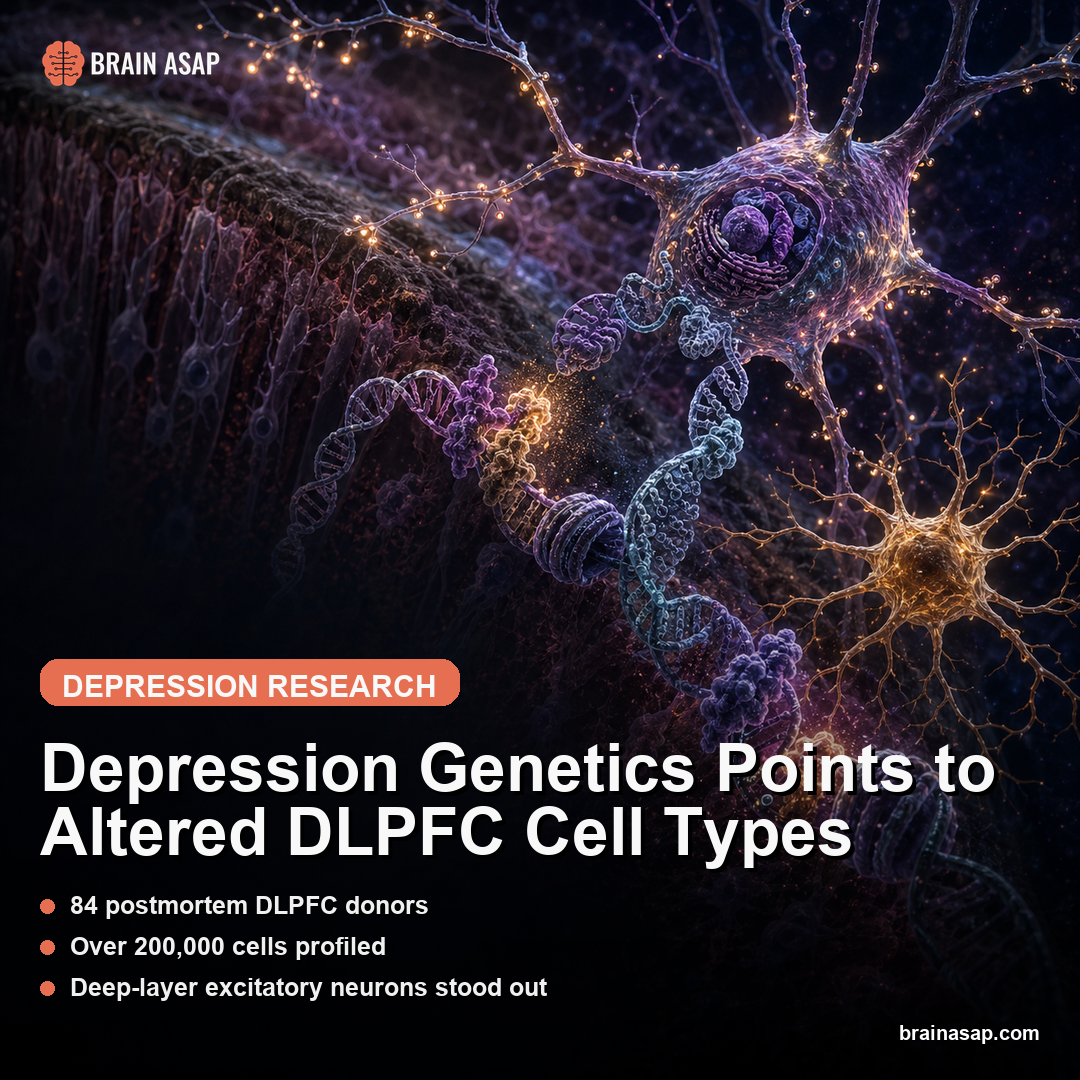
Depression Genetics Points to Altered DLPFC Cell Types
TL;DR: A Nature Genetics study mapped more than 200,000 cells from the dorsolateral prefrontal cortex and found depression-linked regulatory changes concentrated in deep-layer excitatory neurons and a microglia subtype.
Key Findings
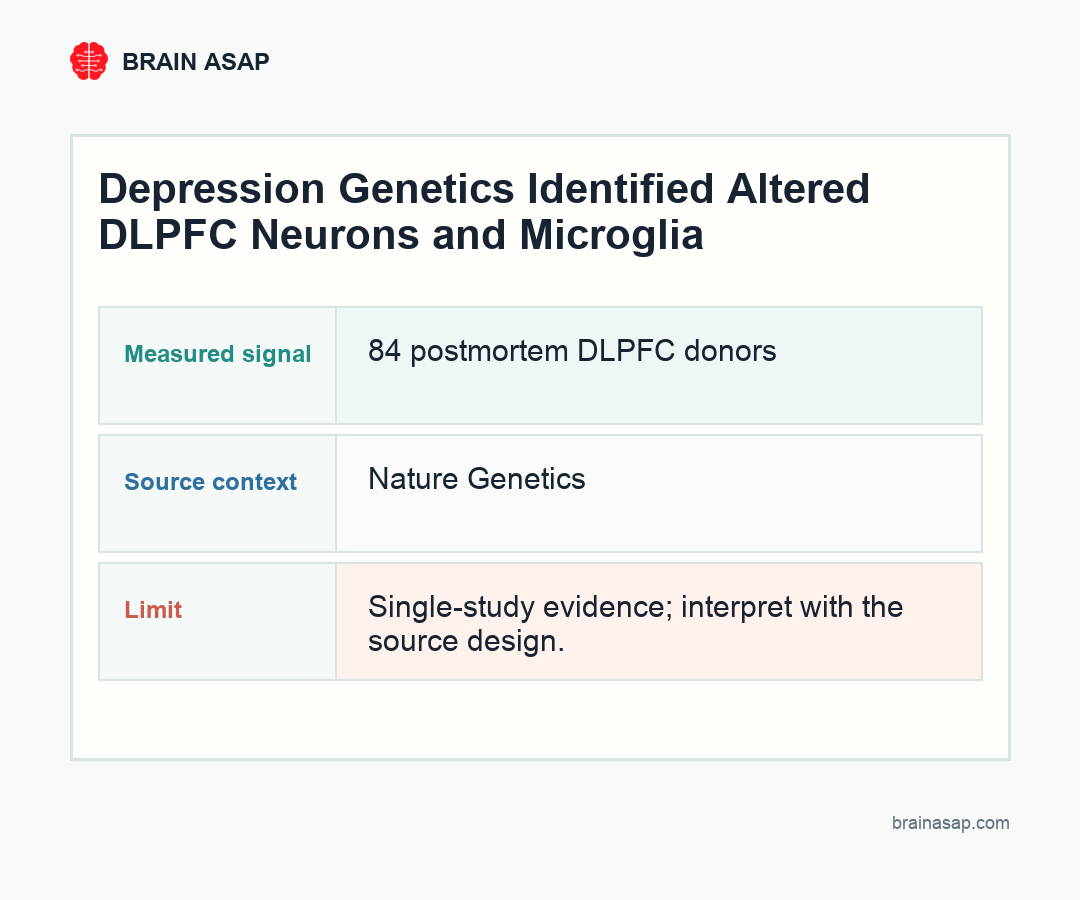
- 84 postmortem DLPFC donors: The analysis compared dorsolateral prefrontal cortex tissue from people with major depression and neurotypical controls.
- Over 200,000 cells profiled: The study combined single-nucleus chromatin accessibility with gene-expression data to identify cell-type-specific regulatory changes.
- Deep-layer excitatory neurons stood out: MDD-associated accessibility changes were prominent in neurons marked by NR4A2 transcription factor motif accessibility.
- Genetic risk landed in those neurons: Depression-associated variants were enriched in the same neuronal regulatory regions, with predicted effects on synaptic communication genes.
- Microglia showed immune-homeostasis changes: A gray matter microglia cluster showed decreased accessibility at sites bound by transcription factors involved in immune regulation.
Source: Nature Genetics (2025) | Chawla et al.
Major depression is often described through symptoms: low mood, anhedonia, sleep disruption, cognitive drag. This paper starts inside the regulatory switches that decide which genes can be read in specific brain cells, then maps where depression genetics may be acting in the dorsolateral prefrontal cortex.
The DLPFC Was Treated as a Cellular Ecosystem
The dorsolateral prefrontal cortex, or DLPFC, is a high-value region for depression research because it helps support cognitive control, emotion regulation, and flexible behavior.
But a piece of cortex is not one thing. It contains excitatory neurons, inhibitory neurons, oligodendrocytes, astrocytes, microglia, endothelial cells, and many subtypes within those categories.
The study used single-nucleus methods to avoid averaging all of that biology into one gray smear. By looking at chromatin accessibility, the team asked which regulatory regions of DNA were open or closed in each cell type. By adding gene expression, they could connect those regulatory states to the genes cells were actually using.
That is the central strength of the paper: it moves depression genetics from a list of risk variants toward specific cell populations in a specific cortical region.
Depression symptoms probably do not arise from one uniform prefrontal defect. A change in deep-layer excitatory neurons can affect long-range cortical output, while a change in microglia can alter synapse pruning, immune signaling, and local tissue state. Those are different intervention and measurement problems.
NR4A2 Marked a Stress-Reactive Neuron Signal
The most striking neuronal signal came from deep-layer excitatory neurons. These cells showed depression-associated changes in chromatin accessibility involving transcription factor motifs, especially NR4A2, an activity-dependent transcription factor that responds to stress biology.
That does not show NR4A2 is the single master switch for depression. Brain disorders rarely give us that kind of clean explanation. But it does suggest a plausible bridge: stress-responsive regulatory programs in cortical excitatory neurons may alter how synaptic genes are controlled in people with major depression.
The same neuronal population was also enriched for MDD-associated genetic variants. That overlap is important because many psychiatric risk variants sit outside protein-coding genes. They often live in regulatory DNA, where the question becomes: regulatory in what cell, at what time, and for which downstream genes?
- Cell type: deep-layer excitatory neurons carried a prominent MDD-linked accessibility signal.
- Regulatory program: NR4A2 motif accessibility pointed toward activity- and stress-responsive gene control.
- Genetic link: depression-associated variants were enriched in the same regulatory neighborhoods.
Microglia Added an Immune-Regulation Layer
The study did not stop at neurons. A gray matter microglia cluster showed decreased chromatin accessibility in people with MDD at binding sites for transcription factors known to regulate immune homeostasis.
Microglia are the brain’s resident immune cells, but that label undersells them. They prune synapses, respond to injury, release signaling molecules, and tune local tissue environments. A microglial regulatory shift in depression is therefore not a side note; it is a clue that immune-state control inside the brain may intersect with mood-circuit biology.
This fits a broader trend in depression research. The field has moved beyond a simple neurotransmitter shortage model. Serotonin, dopamine, and norepinephrine still matter, but so do stress hormones, inflammation, synaptic remodeling, glial cells, and gene regulation.
Genetic Variants Became Functional Hypotheses
A frustrating feature of psychiatric genetics is that large genome-wide association studies can identify risk loci without explaining mechanism. A variant can be statistically real and biologically opaque.
This paper used sequence-based accessibility predictions, donor-specific genotypes, and cell-based assays to push some of those variants toward function. The question was not just whether a variant was near a depression-associated locus. It was whether the variant could plausibly change transcription factor binding and chromatin accessibility in the cell types implicated by the study.
The work is useful even before it becomes therapeutic because it produces testable hypotheses about which regulatory switches might matter most and which cell types deserve deeper experiments.
Postmortem DLPFC Tissue Still Leaves Causality Open
The study used postmortem tissue, so it captures a late-life snapshot of people who had depression, not a movie of how depression developed. Medication history, suicide, comorbidities, and life stress can all complicate interpretation in human brain-bank studies. The authors’ multimodal approach helps, but it cannot erase those limits.
Still, the finding is valuable because it narrows the search space. Instead of saying depression changes the prefrontal cortex, the paper points to deep-layer excitatory neurons with NR4A2-linked regulatory changes and a microglia subtype with altered immune-homeostasis accessibility.
Depression biology is becoming less generic. A future target may not be a whole-neurotransmitter move like “raise serotonin”; it may be a regulatory program in a particular cortical cell type that changes how stress, synaptic communication, and immune signaling meet.
That kind of target is harder to develop, but it is also more biologically honest. A drug, stimulation protocol, or gene-regulation strategy would need to affect the relevant cell state without disrupting every prefrontal circuit that uses the same neurotransmitters.
Chromatin Accessibility Narrowed the Depression Gene List
A gene list can be useful, but it rarely tells the whole depression signal. The same gene can behave differently in different cells, and a risk variant can matter only when it changes a regulatory element that a particular cell type actually uses.
Chromatin accessibility gets closer to that regulatory layer. Open chromatin marks regions where transcription factors can bind and influence gene activity.
Closed chromatin is less available. By mapping those states one nucleus at a time, the authors could ask whether depression-associated regulatory changes were concentrated in neurons, glia, or other cortical cell types.
This is why the NR4A2 and microglia findings are more informative than a generic prefrontal-cortex result. They point toward distinct biological lanes: activity- and stress-responsive excitatory neuron regulation on one side, immune-homeostasis regulation on the other. A future therapy would need that kind of specificity if it hopes to move beyond blunt neurotransmitter-wide effects.
Postmortem Tissue Still Carries Clinical Ambiguity
The study’s strength is also its constraint. Human postmortem DLPFC tissue is precious because it gives direct access to the brain region and cell types of interest. But it cannot separate cause, consequence, treatment history, life history, and terminal state with the certainty of an experiment.
The next step is not to declare a depression mechanism solved. It is to test the candidate regulatory programs in models that can manipulate NR4A2-linked accessibility, excitatory neuron function, microglia state, and stress exposure.
The paper gives the field a better map. It does not yet prove the route.
Paper: Single-nucleus chromatin accessibility profiling identifies cell types and functional variants contributing to major depression. Nature Genetics. 2025;57:1890-1904. DOI: 10.1038/s41588-025-02249-4
Authors: Chawla et al.
Study Design: Postmortem human DLPFC single-nucleus chromatin accessibility and gene-expression study.
Sample Size: 84 individuals and more than 200,000 profiled cells.
Key Statistic: MDD-associated regulatory changes were concentrated in deep-layer excitatory neurons and a gray matter microglia cluster.