Childhood Trauma Made Teen Depression Networks More Random
TL;DR: In 343 adolescents with major depression, childhood trauma was linked to less efficient brain-network organization, partial normalization after treatment, and an fMRI-based model that predicted antidepressant response with 82% balanced accuracy.
Key Findings
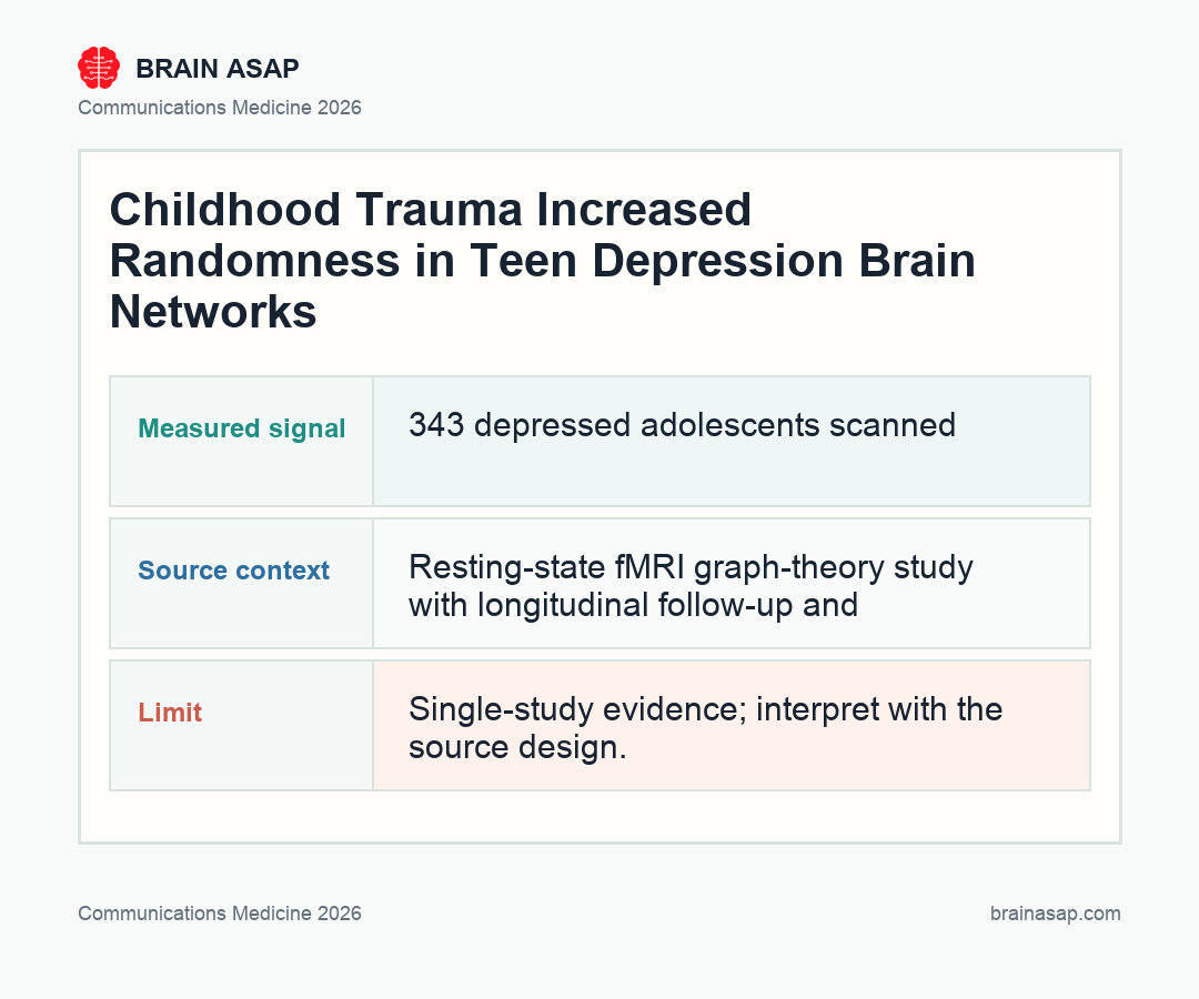
- 343 depressed adolescents scanned: The study analyzed resting-state functional MRI (fMRI) in adolescents ages 10 to 18 with major depressive disorder, including 211 with childhood trauma, 106 without childhood trauma, and 149 healthy controls.
- Trauma shifted DMN hubs: Childhood trauma was linked to disrupted graph-theory measures in default mode network regions including the parahippocampal gyrus, posterior cingulate, and temporal pole.
- Networks looked more random: Depressed adolescents showed higher normalized characteristic path length, a graph-theory signal of less efficient information transfer across the functional connectome.
- Treatment moved some hubs back: In the 71 adolescents with follow-up MRI after treatment, abnormalities partly normalized, especially in the left precuneus and amygdala.
- Baseline fMRI predicted response: A graph convolutional network plus support vector machine model predicted antidepressant response with 82% balanced accuracy, 74% sensitivity, 88% specificity, and AUC 0.90.
Source: Communications Medicine (2026) | Zhu et al.
Depression in a teenager with a trauma history is not just depression plus a bad backstory. In a new adolescent major depressive disorder study, the brain’s communication map looked measurably different when childhood trauma was part of the picture, especially in hubs tied to memory, self-related thought, and emotion.
Why Trauma-Linked Depression Is Not One Brain State
Childhood trauma is one of the strongest risk factors for adolescent depression, but psychiatry has never had a clean biological answer for what that risk does inside the brain. Two teenagers can meet the same diagnostic criteria for major depression while carrying very different developmental histories.
Zhu and colleagues treated that difference as the central question. Instead of asking only whether depressed teens differ from healthy controls, they asked whether depression with childhood trauma has a distinct functional-connectome signature, and whether that signature changes when symptoms improve.
The answer was yes, with boundaries. The study links trauma history to a network pattern rather than establishing causality, and fMRI is not ready for routine clinic use. But it does show that trauma history can carve a measurable pattern into adolescent depression networks, and that those networks are not fixed.
Graph Theory Turned 90 Brain Regions Into a Depression Map
The team used resting-state fMRI, which measures synchronized activity while a person is not performing a task. They converted each scan into a functional network, then applied graph theory, a mathematical way of describing how efficiently a network is organized.
In a healthy brain network, information can move through local clusters while still reaching distant hubs efficiently. In this paper, depressed adolescents showed a higher normalized characteristic path length, often written as lambda. Plainly: the network looked less efficient and more randomly organized.
The trauma-specific pattern landed in regions that make intuitive sense. The default mode network, including the parahippocampal gyrus, posterior cingulate, and temporal pole, helps support autobiographical memory, self-referential thought, and internal simulation. Those are exactly the mental spaces where trauma and depression can become entangled.
The Childhood-Trauma Group Carried More Hub-Level Disruption
The study separated depressed adolescents into those with and without childhood trauma, based on Childhood Trauma Questionnaire cutoffs. The MDD-with-trauma group showed broader nodal abnormalities than the non-trauma depression group, including changes in right parahippocampal gyrus, left cingulate gyrus, and left temporal pole.
Some of those network measures also tracked trauma severity. Sexual abuse scores correlated with nodal degree in the left posterior cingulate and left temporal pole, while physical abuse scores correlated with nodal degree in the left pallidum. These are not large enough to become individual diagnostic markers, but they matter because they connect a clinical history to a specific network architecture.
That distinction is clinically important. If trauma-linked depression has different network features, then lumping all adolescent depression together may blur the very signals that predict treatment response. The paper’s strongest contribution is not that it found a “depression spot.” It found a trauma-conditioned depression network.
8 Weeks of Treatment Partly Reorganized the Connectome
The longitudinal part of the study is what keeps it from being just another single-timepoint imaging snapshot. Treatment outcomes were available for 232 patients, and 71 had repeat MRI after about 8 weeks of standardized antidepressant treatment.
After treatment, some of the network differences that separated depressed adolescents from healthy controls were no longer statistically significant. The movement showed up at two levels:
- Global topology: normalized characteristic path length decreased after treatment.
- Specific hubs: the left amygdala and left precuneus showed notable treatment-linked changes.
- Clinical link: precuneus efficiency tracked symptom change in a subgroup.
The left precuneus result is especially interesting because changes in its nodal efficiency tracked changes in both depression and anxiety scores in a subgroup. The precuneus sits inside the default mode network and is deeply involved in self-processing and internally directed attention. If depression recovery involves changing how the brain handles self-related information, this is a plausible place to see it.
An 82% Response Model Is Promising, Not Clinic-Ready
The paper also tested whether baseline functional connectivity could predict who would respond to antidepressant treatment. The best model combined a graph convolutional network with a support vector machine and reached 82% balanced accuracy, with 74% sensitivity, 88% specificity, and AUC 0.90.
Those are attention-grabbing numbers, but they need restraint. Machine-learning models in neuroimaging can overfit, especially when sample sizes are modest and data come from a single research ecosystem. The authors used cross-validation and stability checks, but the result still needs independent replication before anyone should treat it as a clinical decision tool.
Still, the direction is exactly where depression research needs to go. A teenager who has already been shaped by trauma should not have to cycle blindly through treatments while clinicians wait months to learn whether the first choice worked. The long-term hope is not a brain scan that “diagnoses depression.” It is a brain scan that helps clinicians choose faster and waste less time.
What This Adds to Teen Depression Treatment
The practical message is that childhood trauma may mark a biologically meaningful subtype of adolescent depression. In this study, trauma history was tied to less efficient functional-network organization, especially in default mode and limbic regions, and those abnormalities partly shifted with treatment.
That does not show trauma-linked depression is destiny. If anything, the follow-up MRI results argue the opposite.
The network was disturbed, but not frozen. Symptom improvement came with measurable movement in the brain’s communication map.
The study also makes a subtler point about precision psychiatry. The useful biomarker does not have to be a single molecule, region, or symptom score.
It may be a pattern: a network-level readout that captures how trauma, depression severity, and treatment response intersect. For adolescent depression, that is a much more realistic target than pretending one blood test or one brain region will explain everything.
The next step is replication across sites, scanners, treatment types, and more diverse adolescent populations. But this paper earns attention because it connects three things psychiatry often studies separately: trauma history, brain-network topology, and treatment response. In teenagers, those may be one linked pattern.
Paper: Graph theory reveals functional connectome disruptions in adolescent major depressive disorder with childhood trauma. Communications Medicine. 2026. DOI: 10.1038/s43856-026-01593-8
Authors: Zhu et al.
Study Design: Resting-state fMRI graph-theory study with longitudinal follow-up and machine-learning treatment-response prediction.
Sample Size: 343 adolescents with major depressive disorder, 149 healthy controls, 232 treatment-outcome records, and 71 follow-up MRI scans.
Key Statistic: Baseline functional connectivity predicted antidepressant response with 82% balanced accuracy, 74% sensitivity, 88% specificity, and AUC 0.90.