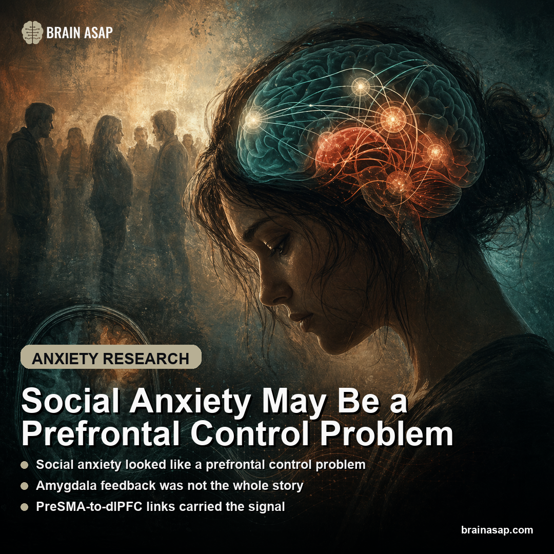
TL;DR: A 2026 study in The British Journal of Psychiatry found that social anxiety disorder was linked to altered communication inside prefrontal control circuits during a functional MRI emotion-regulation task.
Key Findings
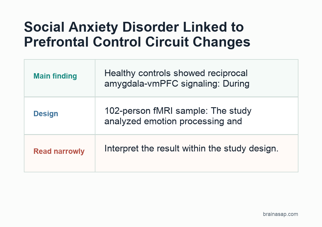
- 102-person fMRI sample: The study analyzed emotion processing and regulation in 61 people with social anxiety disorder and 41 healthy controls using a face-viewing and downregulation task during functional MRI.
- Directed connectivity, not just correlation: Researchers used dynamic causal modeling to estimate the direction of influence among the amygdala, ventromedial prefrontal cortex, dorsolateral prefrontal cortex and pre-supplementary motor area.
- Healthy controls showed reciprocal amygdala-vmPFC signaling: During negative-face observation, controls displayed increased amygdala-to-vmPFC connectivity along with inhibitory vmPFC-to-amygdala connectivity, a result consistent with active feedback regulation.
- Social anxiety differences stayed inside the prefrontal network: Patients did not significantly differ from controls in amygdala-prefrontal coupling; instead, abnormalities appeared in intra-prefrontal links, including inhibitory preSMA-to-dlPFC communication during observation.
- Emotion regulation shifted preSMA-vmPFC dynamics: During deliberate downregulation, social anxiety disorder was marked by bidirectional excitatory connectivity between preSMA and vmPFC, suggesting a different control strategy or compensatory loop.
Source: The British Journal of Psychiatry (2026) | Schrammen et al.
Functional MRI, or fMRI, tracks blood-flow changes as a proxy for brain activity. In this study, it let researchers test how emotion-regulation circuits communicated during the task.
Most brain models of social anxiety tell a familiar story: the amygdala overreacts and the prefrontal cortex fails to rein it in.
This result complicates that model by showing that the clearest differences were concentrated inside the prefrontal network itself rather than in the classic amygdala-versus-cortex battle line.
Prefrontal Control, Not Amygdala Overdrive, Carried the Social-Anxiety Signal
Social anxiety disorder is usually narrated as a limbic alarm problem. The amygdala responds too strongly to social threat, the prefrontal cortex fails to quiet it down, and anxious self-consciousness follows.
That model has been useful, but it also risks being too simple. Functional connectivity studies often show that certain regions correlate differently in patients, yet those analyses cannot say which region is driving which.
Researchers used dynamic causal modeling, a method built to estimate directed influences rather than mere co-activation, to ask a sharper question: when people with social anxiety try to regulate emotion, is the core problem really a broken amygdala-prefrontal line, or is the dysfunction happening somewhere else in the control network?
The participants performed a task with two essential modes:
- Observation: they viewed neutral or negative faces.
- Downregulation: they used a self-chosen strategy to reduce their emotional response.
- Directed modeling: the analysis estimated how control regions influenced one another during each mode.
Social anxiety is not just about passive reactivity. It is also about what happens when people try, and often struggle, to gain control over their internal state in socially charged situations.
The expected culprit did not dominate. Patients with social anxiety disorder did not significantly differ from controls in core amygdala-prefrontal effective connectivity.
The stronger differences were found within the prefrontal cortex itself.
How Healthy Brains Balanced Amygdala Signals With vmPFC Feedback
Before getting to the disorder findings, the control model is worth spelling out because it is more nuanced than the old top-down cartoon.
In healthy participants, viewing negative faces increased amygdala-to-ventromedial prefrontal cortex connectivity while the vmPFC exerted inhibitory influence back onto the amygdala.
That reciprocal loop fits a brain that is not just suppressing emotion but actively monitoring, valuing and reshaping it in real time.
During explicit emotion regulation, healthy controls showed negative modulation from the amygdala to all modeled prefrontal regions. In practical terms, threat processing changed when regulation was engaged, and the prefrontal network adjusted with it.
That is a richer picture than “the cortex turns the amygdala off.” It suggests ongoing coordination between salience detection and control systems.
The control result gives the disorder comparison teeth. If healthy emotion regulation depends on reciprocal and context-sensitive feedback, then pathology can arise from several different points in the loop.
The result does not have to mean the amygdala is hyperactive in every relevant sense.
The broader contribution is to push social anxiety neuroscience toward these circuit-level distinctions. Once researchers stop treating the prefrontal cortex as one undifferentiated boss region, the disorder starts to look more specific and potentially more treatable.
PreSMA-to-dlPFC and preSMA-vmPFC Links Stood Out in Social Anxiety
The prefrontal differences centered on the pre-supplementary motor area, or preSMA, a region involved in control, conflict monitoring and action selection.
During simple observation of emotional faces, patients with social anxiety showed inhibitory connectivity from preSMA to the dorsolateral prefrontal cortex. During active emotion regulation, they showed bidirectional excitatory coupling between preSMA and vmPFC.
This result suggests the disorder involves an altered control architecture rather than a single failed brake on the amygdala.
The preSMA is often discussed in terms of preparing or withholding action, but in social anxiety those “actions” include internal ones: shifting attention, choosing a reappraisal strategy, suppressing avoidance impulses or getting stuck in self-monitoring loops.
If preSMA is pushing differently on dlPFC during emotional observation, patients appear to enter social threat states with a control system that is already configured differently before deliberate regulation even begins.
Then, when they try to regulate, the stronger bidirectional loop with vmPFC likely reflects a more effortful, recursive or unstable valuation process.
This interpretation also fits clinical experience. Socially anxious people often do not simply feel too much fear.
They over-monitor, over-correct and become trapped in self-focused regulatory effort that backfires. A dysregulated prefrontal circuit is a plausible neural mirror of that experience.
What This Means for Biomarkers and Therapy Design
Cognitive behavioral therapy for social anxiety depends heavily on emotion regulation: shifting attention outward, reappraising threat, tolerating uncertainty and interrupting avoidance.
If the disorder’s neural bottleneck sits in intra-prefrontal coordination, then treatment response depends on whether those control circuits can be re-tuned rather than on “calming the amygdala” in a generic sense.
The finding points to three practical research directions:
- Biomarker development: directed connectivity can capture group-relevant information better than simple activation strength.
- Therapy tracking: CBT studies can test whether successful treatment normalizes preSMA, dlPFC and vmPFC coordination.
- Stimulation targets: brain-stimulation studies can focus on control-circuit flexibility rather than only amygdala reactivity.
The study’s leave-one-out cross-validation suggests that effective-connectivity patterns contained enough diagnostic information to predict group membership.
That is far from a clinic-ready test, but it hints that directed circuit measures are designed to capture more than simpler activation contrasts when trying to capture meaningful individual differences.
If preSMA-vmPFC dynamics are part of the problem, future work can test whether successful therapy normalizes those links, or whether brain stimulation aimed at control circuits improves regulatory flexibility.
Even if that work is distant, the study gives researchers a better target map than the old fear-center narrative.
It also offers a psychological payoff. Patients often experience social anxiety as exhausting mental over-control rather than as raw panic alone.
A prefrontal-network account matches that lived phenomenology better than a model built solely around hyperreactive threat detection.
The Findings Are Stronger as a Mechanistic Clue Than a Diagnostic Tool
The study is careful and modern, but there are still limits. A 102-person sample is respectable for a directed-connectivity fMRI study, yet still small relative to the heterogeneity of social anxiety disorder.
Different regulatory strategies, illness durations and comorbid traits can all influence the circuit result.
There is also the usual concern that sophisticated connectivity models can look cleaner than the data truly are.
Dynamic causal modeling is powerful, but it rests on assumptions about model structure and region selection. A different node set may reveal additional pieces of the regulatory network.
Most importantly, the paper does not say the amygdala is irrelevant. It says the decisive difference between patients and controls in this task was not where many people expected to find it.
That is a useful corrective. It nudges the field away from a tired “overactive fear center” explanation and toward a more distributed account of how regulation goes wrong.
The shift can prove more than semantic. If social anxiety is partly a disorder of how prefrontal control nodes coordinate under emotional load, then the next generation of therapy biomarkers, cognitive training and stimulation studies should look there first.
Citation: DOI: 10.1192/bjp.2026.10628. Schrammen et al. Altered prefrontal effective connectivity during emotion regulation in social anxiety disorder. The British Journal of Psychiatry. 2026;1-9.
Study Design: Human fMRI effective-connectivity study using an emotional face-viewing and downregulation task.
Sample Size: 102 participants: 61 people with social anxiety disorder and 41 healthy controls.
Key Statistic: Dynamic causal modeling showed that group differences concentrated in prefrontal effective-connectivity patterns rather than a simple amygdala-prefrontal coupling deficit.
Caveat: The study is mechanistic and imaging-based; it does not prove a diagnostic test or show that changing these circuits will treat social anxiety.