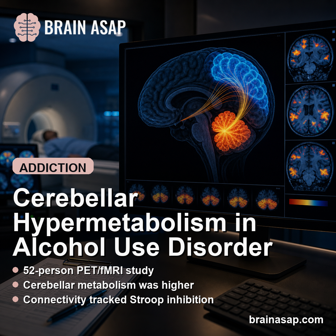
TL;DR: A 2026 study in Translational Psychiatry combined positron emission tomography (PET) and functional MRI (fMRI) in alcohol use disorder and linked cerebellar hypermetabolism to disrupted fronto-cerebellar connectivity and poorer inhibition performance.
Key Findings
- AUD-control sample: Researchers compared 27 people with severe alcohol use disorder (AUD) and 25 healthy controls.
- Multimodal imaging used: The study combined fluorodeoxyglucose PET (FDG-PET) glucose metabolism, resting-state fMRI connectivity, MRI thalamic grey-matter volume, and executive tests.
- Cerebellar metabolism was higher: Alcohol use disorder patients showed higher FDG metabolism in cerebellar lobule VIII than controls, with p = .006.
- Frontal connectivity changed: AUD patients showed stronger negative resting-state functional connectivity between cerebellar lobule VIII and the left superior frontal gyrus.
- Path model favored an indirect route: Cerebellar hypermetabolism related to Stroop inhibition performance through fronto-cerebellar connectivity, with beta = .23 and p = .008.
Source: Translational Psychiatry (2026) | Ritz et al.
The cerebellum is often discussed as a movement structure, but it is also part of loops that support cognition. In alcohol use disorder, those loops can involve the cerebellum, thalamus, and frontal cortex, all of which can affect executive functions such as inhibition and mental flexibility.
The analysis tested whether PET-measured cerebellar hypermetabolism and fMRI-measured cerebellar connectivity captured different aspects of the same fronto-cerebellar circuit abnormality.
27 AUD Patients and 25 Controls Completed PET, fMRI, MRI, and Executive Testing
The sample included 27 patients with severe AUD without Korsakoff syndrome and 25 healthy controls matched on sex, age, and education. AUD patients were assessed during inpatient withdrawal treatment, but none had physical withdrawal symptoms at inclusion.
Every participant underwent several measurements that captured different parts of the same circuit question:
- FDG-PET: A glucose-metabolism scan used to measure cerebellar metabolic activity.
- Resting-state fMRI: A functional connectivity scan used to estimate synchronization between brain regions at rest.
- Structural MRI: A grey-matter measurement used to quantify thalamic volume.
- Executive tests: Trail Making, Stroop, and Modified Card Sorting tasks measured flexibility, inhibition, and set-shifting.
The central seed region was left cerebellar lobule VIII, chosen from prior work showing alcohol-related hypermetabolism in that cerebellar location. The analysis then tested how this seed connected with grey-matter voxels across the brain.
Cerebellar Lobule VIII Had Higher Metabolism in Alcohol Use Disorder
FDG-PET showed higher metabolism in cerebellar lobule VIII in the AUD group than in controls. The reported mean was 1.42 in AUD patients versus 1.25 in healthy controls, with p = .006 and a large effect size.
The thalamus also differed structurally. Thalamic grey-matter volume was lower in AUD patients, with a mean of 0.27 compared with 0.31 in controls and p < .001.
On executive testing, AUD patients were slower on Trail Making Test switching time. Stroop inhibition time showed a trend toward slower performance in AUD patients, and within the AUD group, worse Stroop time correlated with greater cerebellar hypermetabolism and smaller thalamic grey-matter volume.
Fronto-Cerebellar Anticorrelation Was Stronger in AUD Patients
Resting-state fMRI showed that AUD patients had altered connectivity from the cerebellar seed. One finding involved stronger positive connectivity with the left parietal gyrus.
The more clinically central finding involved the left superior frontal gyrus.
Compared with controls, AUD patients showed stronger negative resting-state functional connectivity between cerebellar lobule VIII and the left superior frontal gyrus. In plain terms, higher signal in the cerebellar seed was more strongly paired with lower signal in that frontal region.
The superior frontal region belongs to executive-control circuitry. For inhibition tasks such as the Stroop test, organized anticorrelation may help separate competing brain processes rather than simply represent a broken connection.
- Positive connectivity result: AUD patients showed higher connectivity between cerebellar lobule VIII and the left parietal gyrus.
- Negative connectivity result: AUD patients showed stronger negative connectivity between cerebellar lobule VIII and the left superior frontal gyrus.
- Executive link: In AUD patients, the negative fronto-cerebellar connectivity measure correlated with Stroop inhibition time.
Path Analysis Linked Metabolism, Connectivity, and Stroop Performance
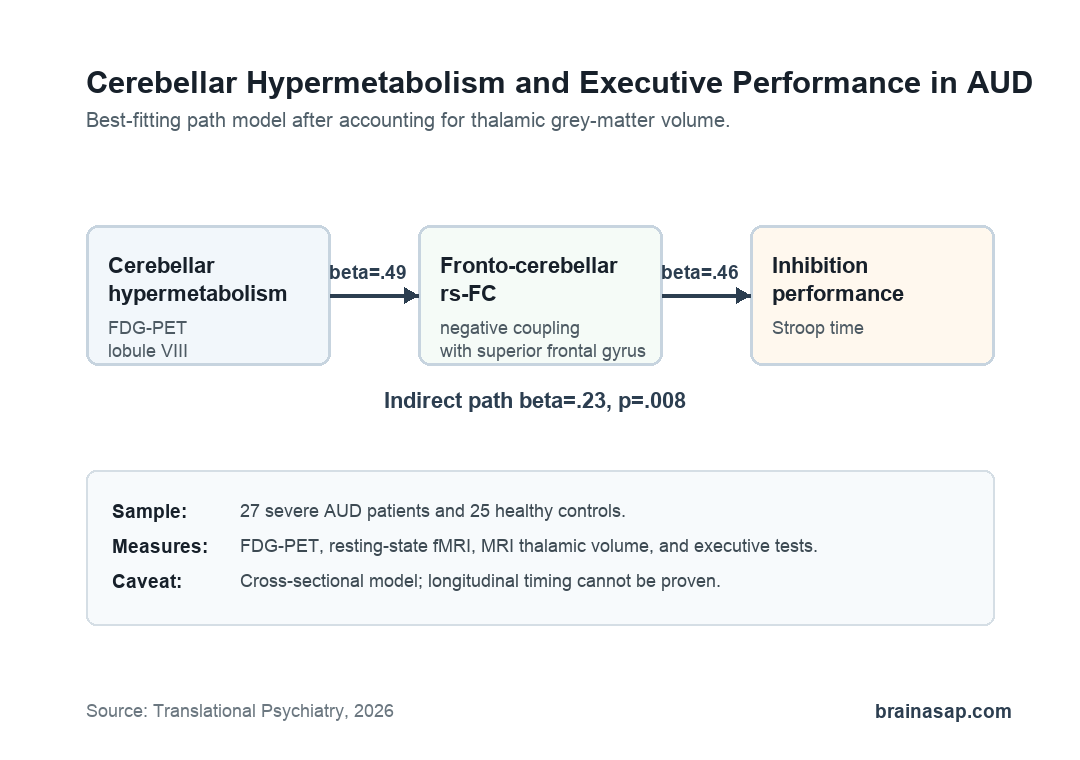
The path analysis tested 3 models in AUD patients. The best-fitting model placed fronto-cerebellar resting-state functional connectivity between cerebellar hypermetabolism and inhibition performance, while controlling for thalamic grey-matter abnormalities.
In that model, cerebellar hypermetabolism was related to fronto-cerebellar connectivity with beta = .49. Fronto-cerebellar connectivity was related to inhibition performance with beta = .46.
The indirect pathway from cerebellar hypermetabolism to inhibition through connectivity was beta = .23, with p = .008.
The interpretation is circuit-specific. Higher cerebellar hypermetabolism was associated with connectivity closer to zero, meaning weaker negative fronto-cerebellar coupling. Weaker negative coupling was associated with worse Stroop inhibition performance.
- Metabolic component: FDG-PET suggested abnormal cerebellar synaptic activity in lobule VIII.
- Connectivity component: Resting-state fMRI suggested altered anticorrelation with the superior frontal gyrus.
- Cognitive component: Stroop time provided the inhibition outcome tied to executive control.
Cross-Sectional Imaging Cannot Prove the Cascade Timing
The study brings PET, fMRI, structural MRI, and cognition into one model. That does not make the model causal.
The design was cross-sectional, so the analysis cannot prove that cerebellar hypermetabolism appears first, then changes connectivity, then impairs inhibition.
Several other limits are important:
- Early abstinence: AUD patients were assessed soon after withdrawal treatment, so short-term abstinence effects cannot be fully excluded.
- Sex imbalance: The AUD group was mostly male, with only 3 women.
- Tobacco exposure: Tobacco use was much more common in AUD patients, although exploratory analyses did not support a major contribution after correction.
- Small imaging sample: The multimodal design was intensive, but the final sample still included only 52 participants.
The cautious takeaway is that cerebellar hypermetabolism may mark alcohol-related brain dysfunction when it weakens organized fronto-cerebellar anticorrelation. Stronger negative connectivity, by contrast, may reflect a compensatory route that supports inhibition performance.
Citation: DOI: 10.1038/s41398-026-04217-w. Ritz et al. Cerebellar hypermetabolism disrupts fronto-cerebellar resting-state functional connectivity and associated executive compensation. Translational Psychiatry. 2026.
Study Design: Cross-sectional multimodal neuroimaging study in alcohol use disorder.
Sample Size: 27 severe AUD patients and 25 healthy controls.
Key Statistic: The best-fitting path model linked cerebellar hypermetabolism to inhibition performance through fronto-cerebellar connectivity, with indirect beta = .23 and p = .008.
Caveat: Cross-sectional imaging cannot establish the temporal sequence of metabolism, connectivity, and executive change.