TL;DR: A 2026 study in Molecular Neurobiology found that Antrodia cinnamomea, a medicinal fungus extract, worked best when carried on citrate-stabilized silver nanoparticles in cell and rat models of Parkinson’s-like dopamine injury.
Key Findings
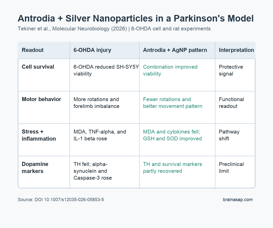
- 6-OHDA created Parkinson’s-like injury: The toxin reduced SH-SY5Y cell viability and produced unilateral dopamine-system damage in rats.
- Combination treatment was strongest: Antrodia cinnamomea plus silver nanoparticles improved cell survival, motor behavior, inflammatory markers, and tissue preservation more consistently than either component alone.
- Oxidative stress and inflammation moved lower: Combination treatment lowered MDA, TNF-alpha, and IL-1 beta while improving antioxidant markers such as GSH and SOD.
- Dopamine-system markers partly recovered: The combination group showed stronger tyrosine hydroxylase immunostaining, a dopamine-neuron marker, and less alpha-synuclein staining than untreated lesioned rats.
- The evidence is preclinical: The rat experiment used a short-term 6-OHDA lesion model in male animals, so it does not prove a treatment benefit in people with Parkinson’s disease.
Source: Tekiner et al. Molecular Neurobiology. 2026.
Parkinson’s disease research often uses toxin models to separate dopamine-system injury from the full complexity of the human disease.
That setup lets researchers ask whether a candidate treatment changes behavior only, or whether it also shifts inflammation, oxidative stress, apoptosis, and dopamine-neuron markers.
The result does not make a supplement ready for Parkinson’s treatment. In a controlled preclinical model, the combined formulation affected several injury pathways at once.
Parkinson’s disease involves more than dopamine loss alone. Oxidative stress, inflammation, apoptosis, alpha-synuclein accumulation, and mitochondrial stress can all contribute to neuron damage.
Because the evidence came from cells and rats, these data are mechanistic. They show a coordinated biological response, not clinical efficacy.
Researchers Tested 6-OHDA Injury in Cells and Male Rats
The experiment began with differentiated SH-SY5Y cells, a human neuroblastoma-derived cell line often used as a dopamine-neuron-like laboratory model. Researchers exposed the cells to 6-hydroxydopamine (6-OHDA), a toxin that creates oxidative stress and dopamine-neuron injury in Parkinson’s disease research.
The animal experiment used 63 male rats divided into 9 experimental groups. Some rats received 6-OHDA to create unilateral nigrostriatal injury, meaning one side of the dopamine pathway from the substantia nigra to the striatum was damaged.
The main treatment groups compared:
- Antrodia cinnamomea: the fungus extract was given orally at 100 mg/kg.
- Silver nanoparticles: citrate-stabilized AgNPs were tested at 1 mg/kg.
- Combination treatment: Antrodia cinnamomea and AgNPs were mixed in a 1:1 ratio before treatment.
- L-DOPA comparator: levodopa was included as a dopamine-replacement reference treatment, not as a disease-modifying control.
Researchers then measured behavior, oxidative-stress markers, inflammatory cytokines, dopamine and acetylcholine by LC-MS/MS, substantia nigra histology, and protein markers tied to neuron survival and apoptosis.
Combination Treatment Improved Parkinson’s-Like Motor Readouts
The 6-OHDA lesion produced expected Parkinson’s-like motor impairment. Lesioned rats showed more apomorphine-induced rotations, less balanced forelimb use, and reduced locomotor activity.
Those behavior tests are indirect but useful in this model. Rotation behavior reflects asymmetry after unilateral dopamine injury.
The cylinder test checks whether rats rely more on one forelimb. Open-field locomotion gives a broader view of movement.
Compared with untreated 6-OHDA rats, treatment groups had fewer rotations and better movement readouts. The Antrodia plus AgNP group was the most consistent across behavior, tissue, and molecular measures.
L-DOPA also reduced some motor impairment, which is expected because levodopa supports dopamine signaling.
The Antrodia-nanoparticle comparison asked a different question: whether the formulation also changed oxidative stress, inflammation, and neuron-preservation markers.
Oxidative Stress and Inflammatory Markers Moved in a Protective Direction
In the 6-OHDA group, malondialdehyde (MDA), a lipid-peroxidation marker, increased. At the same time, antioxidant defenses such as glutathione (GSH) and superoxide dismutase (SOD) were reduced.
The combination treatment shifted the redox profile. MDA decreased, while GSH and SOD activity improved relative to the untreated lesion group.
The inflammatory markers told a similar story. Untreated 6-OHDA rats had higher TNF-alpha and IL-1 beta, 2 inflammatory cytokines that can rise during neuroinflammatory stress.
Antrodia, AgNPs, and especially the combined formulation reduced those cytokine levels compared with untreated lesioned rats.
The result should be read as a pathway signal. Lower inflammatory and oxidative-stress markers suggest the treatment changed the model’s injury environment, but those measures do not prove long-term neuroprotection in human Parkinson’s disease.
Dopamine-Neuron and Apoptosis Markers Also Shifted
Histology showed more severe substantia nigra neuron damage after 6-OHDA. The combination group had less visible tissue injury and better preservation of neuronal morphology.
Immunostaining also favored the combined formulation. Tyrosine hydroxylase (TH), a marker for dopamine-producing neurons, was lower after 6-OHDA and partly restored in treated groups, with the clearest recovery in the combined treatment group.
Alpha-synuclein immunoreactivity increased after the lesion and decreased after combination treatment. The model did not recreate full Lewy body disease, but a protein linked to Parkinson’s pathology moved in the expected injury direction and then partly normalized.
Western blot results added survival-pathway context:
- Caspase-3: this apoptosis-related protein increased after 6-OHDA and was lower in treated groups.
- Bcl-2: this cell-survival protein fell after 6-OHDA and improved most clearly with combination treatment.
- PI3K: this pro-survival signaling marker decreased after injury and shifted upward in treated animals.
- Agmatinase: expression dropped after the lesion and was partly restored by treatment.
Taken together, the molecular profile supports a multitarget interpretation: the formulation did not only change one dopamine readout. It affected oxidative stress, inflammation, apoptosis, and dopamine-neuron markers in the same general direction.
Translation Depends on Safety, Dosing, and Better Parkinson’s Models
The most important boundary is the model. 6-OHDA creates rapid toxin-induced dopamine injury. Human Parkinson’s disease is slower, more heterogeneous, and includes features that this model cannot fully reproduce.
The rat study also used male animals only. Sex differences can affect neuroinflammation, dopamine injury, and treatment response, so female animals will need direct testing before the result can be generalized.
Nanoparticle safety is another constraint. Silver nanoparticles can behave differently depending on size, coating, dose, tissue distribution, and exposure duration.
A beneficial short-term signal in a rodent model does not settle long-term neurotoxicity or pharmacokinetics.
Antrodia extracts also vary by cultivation and extraction method. A reproducible therapeutic claim would require standardized composition, dose-response testing, chronic-model studies, and toxicology.
For now, the study supports a specific preclinical conclusion: Antrodia cinnamomea loaded onto citrate-stabilized silver nanoparticles reduced several 6-OHDA injury signals more consistently than either component alone. That is a useful lead for Parkinson’s mechanism research, not a clinical recommendation.
Citation: DOI: 10.1007/s12035-026-05853-5. Tekiner et al. Modulation of Oxidative Stress and Apoptosis by Antrodia cinnamomea-Loaded Citrate-Stabilized Silver Nanoparticles in Experimental Parkinsonism. Molecular Neurobiology. 2026;63:576.
Study Design: Preclinical SH-SY5Y cell experiment plus unilateral 6-OHDA rat model of Parkinson’s-like dopamine-system injury.
Sample/Model: Differentiated SH-SY5Y cells and 63 male rats divided into 9 experimental groups.
Key Statistic: The combination treatment reduced apomorphine-induced rotations, lowered MDA, TNF-alpha, and IL-1 beta, restored GSH and SOD activity, and improved TH and apoptosis-related protein markers versus untreated 6-OHDA rats.
Caveat: Short-term preclinical toxin-model evidence; the study does not prove clinical benefit or long-term nanoparticle safety in people with Parkinson’s disease.