TL;DR: A 2026 preclinical study in the International Journal of Nanomedicine developed human umbilical cord mesenchymal stem cell membrane-coated PLGA nanoparticles to co-deliver doxorubicin and curcumin to glioblastoma models, improving tumor-cell uptake and mouse tumor suppression without proving human efficacy.
Key Findings
- MSC membrane coating: Researchers coated PLGA nanoparticles with human umbilical cord mesenchymal stem cell membranes to add tumor-homing properties.
- DOX plus CUR: The platform co-loaded doxorubicin (DOX) and curcumin (CUR), aiming to combine chemotherapy potency with toxicity reduction.
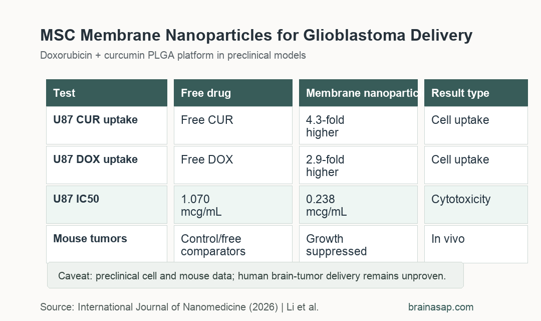
- 4.3-fold CUR uptake: In U87 glioblastoma cells, CUR fluorescence was 4.3-fold higher with CUR/DOX@PLGA-M than free CUR.
- IC50 0.238 micrograms/mL: CUR/DOX@PLGA-M showed stronger U87 cytotoxicity than free DOX, whose IC50 was 1.070 micrograms/mL.
- Mouse tumor suppression: In subcutaneous GL261 glioblastoma models, tail-vein CUR/DOX@PLGA-M treatment suppressed tumor growth while liver and kidney markers stayed in normal ranges.
Source: International Journal of Nanomedicine (2026) | Li et al.
Glioblastoma is an aggressive brain tumor where drug delivery is limited by the blood-brain barrier and the blood-brain tumor barrier. Doxorubicin is potent against cancer cells, but brain delivery and systemic toxicity limit its usefulness.
This study tested a biomimetic delivery idea: wrap drug-loaded polymer nanoparticles in mesenchymal stem cell membrane so the particles inherit some tumor-homing behavior.
MSC Membrane-Coated PLGA Nanoparticles Carried DOX and Curcumin
The platform used poly(lactic-co-glycolic acid) (PLGA), a biodegradable polymer often used for drug delivery. The optimized formulation used a PLGA:curcumin:doxorubicin mass ratio of 150:1:1.
Researchers then coated the nanoparticles with membranes from human umbilical cord mesenchymal stem cells. The final construct was named CUR/DOX@PLGA-M.
- Core carrier: PLGA nanoparticles provided the drug-loaded scaffold.
- Chemotherapy cargo: Doxorubicin supplied the main tumor-killing agent.
- Curcumin cargo: Curcumin was included to enhance anticancer effects and potentially reduce doxorubicin toxicity.
- Membrane camouflage: hUC-MSC membrane coating was intended to improve glioblastoma targeting and reduce nonspecific clearance.
The optimized nanoparticles had reported encapsulation efficiencies of 78.5% for curcumin and 88.5% for doxorubicin. Their hydrodynamic diameter was about 142.1 nm.
Glioblastoma Cell Uptake Increased With the Membrane Coating
The uptake experiments used HCMEC/D3 endothelial cells and U87 glioblastoma cells. Compared with free curcumin, CUR fluorescence in U87 cells was 4.3-fold higher with CUR/DOX@PLGA-M.
Doxorubicin uptake also improved. DOX fluorescence in U87 cells increased by 2.9-fold with the membrane-coated nanoparticle compared with free DOX.
- Endothelial-cell uptake: CUR fluorescence was 3.9-fold higher with CUR/DOX@PLGA-M than free CUR in HCMEC/D3 cells.
- Glioblastoma-cell uptake: CUR fluorescence was 4.3-fold higher and DOX fluorescence was 2.9-fold higher in U87 cells.
- Subcellular pattern: CUR accumulated mainly in cytoplasm, while DOX entered the nucleus.
- BBB model: The paper reported improved transport through an in vitro blood-brain barrier model and better penetration into 3D glioblastoma spheroids.
The barrier experiment used HCMEC/D3 endothelial cells with U87 cells in the lower chamber, while the spheroid experiment tested whether fluorescence reached deeper tumor-cell layers. Those models do not fully reproduce a living brain tumor, but they are more informative than a flat cell-culture uptake test alone.
Curcumin-Doxorubicin Nanoparticles Lowered the U87 IC50
Cell-viability assays suggested that curcumin strengthened doxorubicin’s anti-proliferative effect in U87 glioblastoma cells. Free DOX had an IC50 of 1.070 micrograms/mL.
Adding curcumin lowered the free DOX IC50 to 0.291 micrograms/mL. Encapsulation in CUR/DOX@PLGA-M lowered it further to 0.238 micrograms/mL.
- Free DOX: IC50 was 1.070 micrograms/mL.
- Free DOX plus CUR: IC50 was 0.291 micrograms/mL.
- CUR/DOX@PLGA: IC50 was 0.299 micrograms/mL.
- CUR/DOX@PLGA-M: IC50 was 0.238 micrograms/mL, the strongest reported potency among these comparisons.
The study also reported greater apoptosis and reduced migration, invasion, and angiogenesis markers with the biomimetic platform. Those mechanisms support the tumor-growth result but still come from lab and animal models.
Mouse Tumor Data Supported Antitumor Activity but Used a Subcutaneous Model
The in vivo experiment used C57BL/6 mice with subcutaneous GL261 glioblastoma tumors. Treatments started on day 10 after inoculation and were given by tail-vein injection every 48 hours for 7 doses.
CUR/DOX@PLGA-M suppressed tumor growth more effectively than free drug or uncoated nanoparticle comparisons. Tumor tissues also showed more apoptosis by TUNEL staining.
- Model type: The tumor model was subcutaneous, not an orthotopic brain tumor model.
- Safety readout: Liver markers including ALT, ALB, ALP, and AST stayed within normal physiological ranges.
- Renal readout: Urea and creatinine showed no statistically significant deviations from control values.
The subcutaneous model makes tumor measurement straightforward, but it does not fully recreate the human brain-tumor environment or the intact blood-brain barrier challenge.
Brain Tumor Nanoparticle Results Need Orthotopic and Clinical Testing
The study is best read as a drug-delivery engineering result. It showed that a stem-cell-membrane coating can improve glioblastoma-cell recognition, drug uptake, and antitumor effects in preclinical delivery systems.
The system is not ready for patients with glioblastoma based on these data alone. Human translation would need orthotopic brain-tumor models, dosing and toxicity studies, manufacturing controls, immune-safety testing, and eventually clinical trials.
The most specific next question is whether CUR/DOX@PLGA-M can improve brain tumor drug accumulation and survival in models that reproduce the human brain-tumor barrier more closely.
Citation: DOI: 10.2147/IJN.S571089. Li et al. Mesenchymal Stem Cells Membrane Biomimetic Nanoplatform for Glioblastoma-Targeted Combinatorial Chemotherapy. International Journal of Nanomedicine. 2026;21:571089.
Study Design: Preclinical nanomedicine study using glioblastoma cell assays, blood-brain barrier models, tumor spheroids, and mouse tumor experiments.
Sample/Model: U87 and GL261 glioblastoma models, HCMEC/D3 endothelial cells, and subcutaneous GL261 tumors in C57BL/6 mice.
Key Statistic: CUR/DOX@PLGA-M lowered U87 cell IC50 to 0.238 micrograms/mL, compared with 1.070 micrograms/mL for free doxorubicin.
Caveat: The animal efficacy experiment used a subcutaneous glioblastoma model, so brain-barrier and clinical translation remain unresolved.