TL;DR: A 2026 study in Science Advances reported that disrupting the membrane-associated periodic skeleton (MPS), an actin-spectrin lattice beneath neuron membranes, reduced the fitted amyloid precursor protein (APP) uptake time constant in cultured mouse hippocampal neurons and increased intracellular amyloid-beta 42 under APP-overexpression conditions. The cell-culture experiments do not show that stabilizing the MPS prevents Alzheimer’s disease in animals or people.
Key Findings
- Cultured-neuron model: Researchers studied hippocampal neurons collected from embryonic day 18 CD-1 mouse embryos and matured for 21 to 28 days in culture.
- Four uptake routes: Surface-localized clathrin-, caveolin-, flotillin-, and fast endophilin-mediated pits were preferentially located in MPS-free openings.
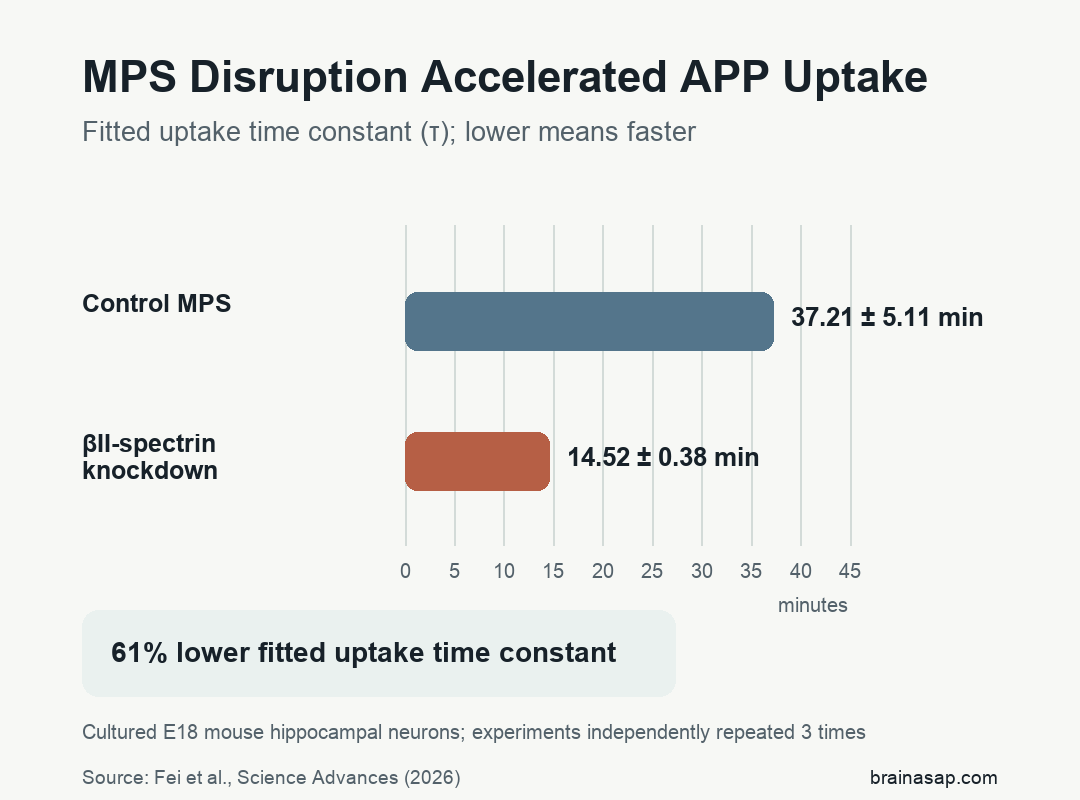
- Faster APP uptake: After beta-II-spectrin knockdown disrupted the MPS, the fitted APP uptake time constant decreased from 37.21 ± 5.11 to 14.52 ± 0.38 minutes, a 61% reduction that indicates faster internalization.
- Alzheimer’s boundary: Aβ42-positive and cleaved-caspase-3 area fractions increased when MPS disruption was combined with engineered APP overexpression, but no living animal or human Alzheimer’s outcome was tested.
Source: Fei et al., Science Advances (2026).
Endocytosis allows a neuron to pull receptors, nutrients, and membrane components into the cell. The membrane-associated periodic skeleton sits just under the membrane, where actin rings connected by spectrin proteins form a repeating lattice with spacing of about 190 nanometers.
The MPS lattice is not a solid shell; small membrane regions without MPS create openings where endocytic pits can form. The experiments tested whether those openings regulate several uptake systems and whether weakening the lattice changes APP processing relevant to Alzheimer’s biology.
Cultured Mouse Neurons Were Tested Across Four Endocytic Pathways
Researchers isolated hippocampi from embryonic day 18 CD-1 mouse embryos, dissociated the tissue into individual cells, and maintained the neurons in culture. Most experiments used mature cultures at 21 to 28 days in vitro, when the MPS is well developed in axons and more established in dendrites.
The team mapped four major endocytic pathways:
- Clathrin-mediated endocytosis: A common route for receptors and nutrients, mapped with clathrin and assessed with transferrin and low-density lipoprotein uptake.
- Caveolin-mediated endocytosis: A lipid-raft–associated route mapped with caveolin-1; ligand-induced activity was assessed using antibody-triggered uptake of overexpressed HA-mGluR5a.
- Flotillin-mediated endocytosis: A clathrin-independent route organized by flotillin proteins.
- Fast endophilin-mediated endocytosis: A rapid receptor-uptake route mapped with endophilin-A2 and assessed using antibody-triggered internalization of endogenous neural cell adhesion molecule 1.
Structured illumination microscopy mapped pit density across the cell. Three-dimensional stochastic optical reconstruction microscopy (STORM), with lateral resolution of 20 to 30 nanometers, then placed individual pits relative to the MPS.
Across three independent repetitions, area-fraction analyses used 20 to 25 imaging fields or regions per condition. Pit-localization methods used 50 to 100 axon or dendrite segments, MPS autocorrelation used 30 to 50 neurite segments, and axonal colocalization analyses used 70 to 130 regions.
The report did not state the total number of embryos, pregnant mice, or independent culture preparations.
Endocytic Pits Occupied Openings in the Membrane Skeleton
All four pit types appeared in the axon initial segment, distal axons, cell body, and dendrites. Their densities differed by compartment: clathrin and flotillin pits were most dense in distal axons, while caveolin and endophilin-A2 pits were denser in dendrites.
STORM images showed that surface-localized clathrin-, caveolin-, flotillin-, and endophilin-A2–marked pits were preferentially located in circular MPS-free openings. Randomized-position controls did not reproduce that pattern.
To test function rather than location alone, researchers used short hairpin RNA to reduce beta-II-spectrin, a core MPS component, by about 70%. Mature neurons with beta-II-spectrin knockdown had more pits across all four pathways in the axon initial segment, distal axons, and dendrites.
Immature seven-day cultures provided a developmental control because their dendritic MPS was not yet established; beta-II-spectrin knockdown did not increase pit density there. That comparison supports MPS disruption, rather than an unrelated effect of losing beta-II-spectrin, as the explanation in mature neurons.
MPS Disruption Accelerated Receptor and APP Uptake
Loss of the lattice changed uptake speed as well as pit density: the fitted transferrin uptake time constant fell from 17.15 ± 0.89 minutes in control neurons to 8.29 ± 0.37 minutes after beta-II-spectrin knockdown. Low-density lipoprotein uptake changed from 54.63 ± 2.45 to 35.25 ± 1.49 minutes.
For APP, researchers attached a pH-sensitive green fluorescent protein variant to the neuronal APP695 isoform and used a fluorescent nanobody against that tag to initiate and track internalization. APP uptake used both clathrin-dependent and clathrin-independent routes under these conditions.
The fitted APP uptake time constant decreased from 37.21 ± 5.11 minutes with an intact MPS to 14.52 ± 0.38 minutes after beta-II-spectrin knockdown. A lower fitted time constant means faster uptake; it is not the exact time required for every APP molecule to enter a neuron.
Endocytosis Activated ERK and Spectrin-Cleaving Proteases
Transferrin uptake and antibody-triggered internalization of overexpressed HA-mGluR5a and endogenous neural cell adhesion molecule 1 increased extracellular signal-regulated kinase (ERK) activity. ERK activation persisted for up to 60 minutes after stimulation.
Blocking endocytosis reduced ERK activation and protected the MPS, while inhibiting MEK, the enzyme directly upstream of ERK, also prevented lattice degradation. These interventions place receptor uptake and ERK activation before MPS breakdown in the experimental sequence.
Western blots identified spectrin fragments produced mainly by calpain, a protein-cutting enzyme, with a smaller contribution from caspases. Calpain or caspase inhibitors preserved more of the MPS and reduced subsequent transferrin, glutamate-receptor, and APP uptake.
Together, the interventions support a proposed culture-model loop: receptor uptake increased ERK signaling, ERK-pathway activity promoted protease-dependent MPS breakdown, and MPS weakening permitted more uptake. Broadly blocking this process would not automatically be beneficial, because normal endocytosis is required for neuronal nutrition, receptor recycling, and communication.
Aβ42 Increased Only Under Engineered APP Conditions
Researchers next overexpressed C-terminal GFP-tagged APP695 in either its normal form or with the Swedish familial Alzheimer’s mutation. Both conditions increased the Aβ42-positive area fraction in somatodendritic regions compared with neurons without APP overexpression.
Combining APP overexpression with beta-II-spectrin knockdown produced higher intracellular Aβ42-positive area fractions in somatodendritic regions and higher cleaved-caspase-3 area fractions in dendrites. Without APP overexpression, Aβ42 area fractions were comparable in control and MPS-disrupted neurons.
Main limitations: These were cultured embryonic mouse neurons, not an intact aging brain. The APP uptake assay used an engineered tag and nanobody, while the amyloid experiments overexpressed C-terminal GFP-tagged APP constructs.
Many denominators were imaging fields or neurite segments rather than animals. Total embryo and culture-batch counts were not reported, although the experiments were repeated three times.
The results therefore identify a cell mechanism, not an Alzheimer’s treatment. Testing whether selective MPS stabilization changes amyloid accumulation, cognition, or safety would require living disease models before any human implication could be evaluated.
Citation: DOI: 10.1126/sciadv.aeb0803. Fei J et al. Membrane-associated periodic skeleton regulates major forms of endocytosis in neurons through a signaling-driven positive feedback loop. Science Advances. 2026;12(7):eaeb0803.
Study Design: Mechanistic cell-culture experiments using super-resolution microscopy, beta-II-spectrin knockdown, ligand-uptake assays, protease inhibitors, and APP overexpression.
Sample/Model: Embryonic day 18 CD-1 mouse hippocampal neurons cultured for 21 to 28 days; experiments were independently repeated three times, with total embryo and culture counts not reported.
Key Statistic: The fitted APP uptake time constant decreased from 37.21 ± 5.11 to 14.52 ± 0.38 minutes after beta-II-spectrin knockdown, indicating faster internalization.
Caveat: Engineered cultured neurons and APP overexpression cannot establish that MPS stabilization prevents amyloid disease or neuronal loss in animals or people.