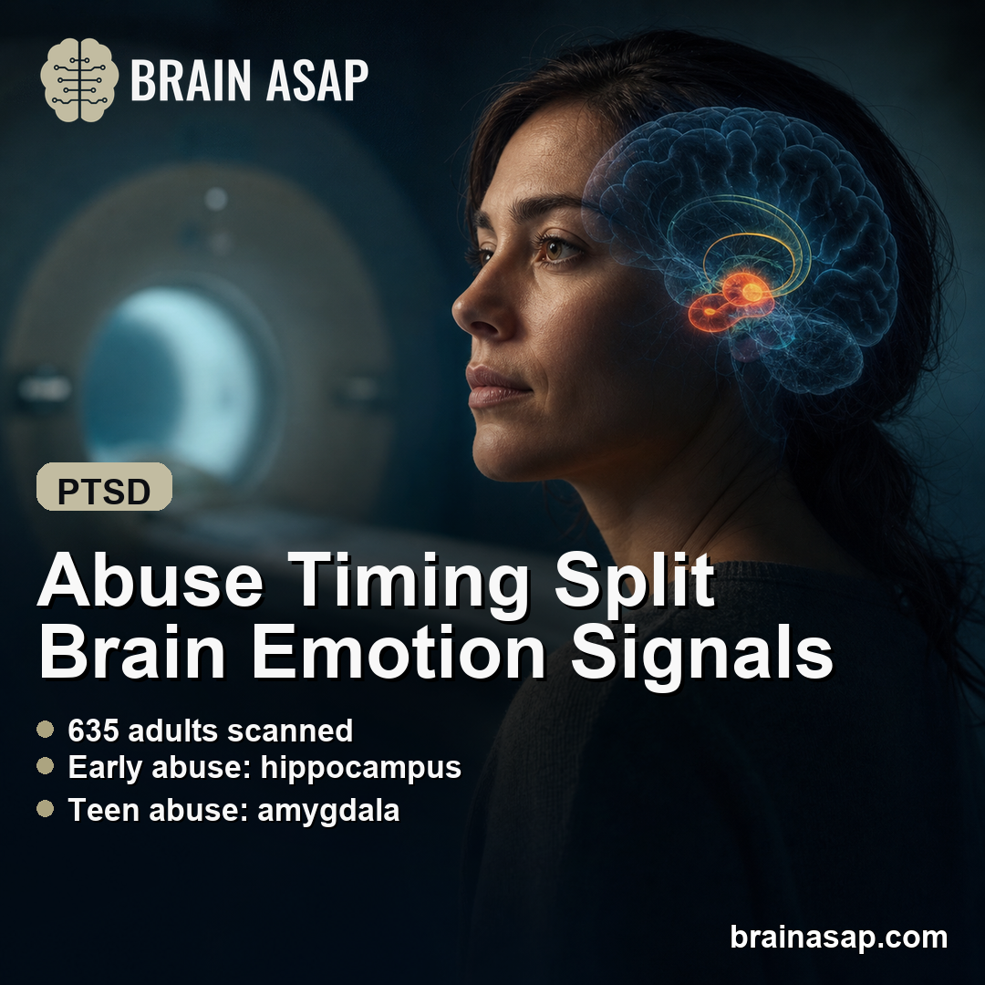
TL;DR: A 2026 fMRI study in Molecular Psychiatry linked abuse before age 13 to stronger hippocampal responses during non-conscious negative emotion processing, while abuse during adolescence was linked to stronger amygdala responses during conscious emotion viewing.
Key Findings
- Timing split the limbic response: The sample included 635 adults after exclusions and quality checks, including people with major depression, anxiety disorders, PTSD, and healthy comparison participants.
- 236 reported abuse or neglect before 18: Researchers compared 399 adults without reported abuse or neglect with adults who reported emotional, physical, or sexual abuse or neglect before adulthood.
- Early abuse tracked hippocampal activity: Abuse before age 13 was linked to stronger right hippocampal activation during non-conscious processing of negative facial emotions.
- Teen abuse tracked amygdala activity: Abuse first reported between ages 13 and 18 was linked to higher amygdala activation during conscious viewing of emotional faces.
- Current symptoms did not explain the main pattern: Depression, anxiety, and stress scores were not correlated with the hippocampal or amygdala activation measures.
Source: Molecular Psychiatry (2026) | Korgaonkar et al.
Childhood abuse is often discussed as one exposure, but timing may matter for how the adult brain handles emotion. In this functional MRI (fMRI) study, the difference was not just whether abuse occurred. The strongest signals separated abuse before age 13 from abuse during adolescence.
The researchers studied adults with and without depression, anxiety disorders, and PTSD, then measured brain responses while participants viewed emotional faces. Some faces were shown long enough for conscious perception. Others were flashed briefly and masked, a task meant to capture non-conscious emotion processing.
Abuse Timing Split the Limbic Brain Signal
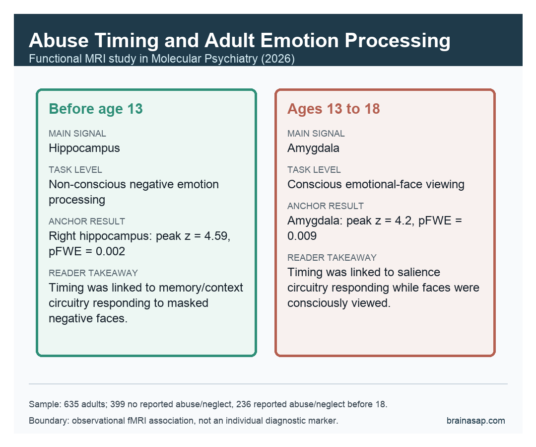
The main result was a timing-specific split across two limbic regions. Adults who reported abuse before age 13 showed stronger right hippocampal activation during non-conscious processing of negative faces. Adults who reported abuse from ages 13 to 18 showed stronger amygdala activation during conscious emotion viewing.
That matters because the hippocampus and amygdala do related but different jobs. The hippocampus helps bind emotion to memory and context. The amygdala helps detect emotionally important stimuli and can become more reactive to threat.
The study does not prove that abuse caused those brain differences. It does show that abuse timing was associated with different emotion-processing patterns later in life, even across diagnostic groups.
The Sample Mixed Diagnoses on Purpose
The analysis included 635 adults after exclusions and fMRI quality checks. There were 399 participants without reported abuse or neglect and 236 participants who reported emotional, physical, or sexual abuse or neglect before age 18.
The diagnostic mix was broad:
- Major depressive disorder: 235 participants.
- Anxiety disorders: 56 participants.
- PTSD: 62 participants.
- Healthy comparison participants: 282 participants, including trauma- and grief-control groups.
This transdiagnostic design is useful because early adversity rarely maps neatly onto one adult diagnosis. The same abuse history can show up in depression, PTSD, anxiety symptoms, or no current disorder. A mixed sample lets researchers ask whether the brain signal cuts across those clinical labels.
Early Abuse Was Linked to Non-Conscious Hippocampal Reactivity
During the non-conscious face task, the full abuse-versus-no-abuse comparison did not show a simple main effect. Instead, there was a group-by-emotion interaction in the right hippocampus, with a peak z-score of 4.48 and family-wise-error corrected p = 0.003.
Post-hoc tests suggested the pattern was driven by stronger hippocampal responses to negative emotions such as anger, disgust, fear, and sadness compared with happy or neutral faces. When the abuse group was split by timing, the hippocampal result remained significant only for adults who reported abuse before age 13.
That does not mean the hippocampus is a trauma detector. A more careful interpretation is that early abuse was associated with altered contextual or memory-linked processing of emotional cues that participants were not consciously identifying.
Adolescent Abuse Was Linked to Conscious Amygdala Reactivity
The conscious face task showed a different timing pattern. Adults who reported abuse between ages 13 and 18 had higher amygdala activation across emotional faces, with a peak z-score of 4.2 and family-wise-error corrected p = 0.009 versus the no-abuse group.
The adolescent-onset group also showed greater amygdala activation than the pre-13 abuse group in a direct comparison. That direct comparison reached a peak z-score of 4.48 and family-wise-error corrected p = 0.002.
The authors interpreted this as evidence that adolescent abuse may be tied more strongly to conscious emotion-processing pathways. The result fits a broader literature in which threat-related adversity is linked to amygdala hyperactivation, but the paper is careful about what the task can and cannot show.
Symptoms and Diagnoses Did Not Fully Account for the Findings
Adults in the abuse group had higher current depression, anxiety, and stress scores, and psychiatric diagnoses were more common in that group. That imbalance is important because it could have explained the fMRI results if the brain signal simply reflected current symptom burden.
The authors tested that concern. They reported that current depression, anxiety, and stress symptoms were not correlated with the hippocampal or amygdala activation measures. The hippocampal result did not show a diagnosis-by-abuse interaction.
The amygdala pattern was more diagnosis-sensitive. Post-hoc analyses suggested the adolescent-abuse amygdala effect was significant mainly in the depression and stress/PTSD groups, not in the anxiety or healthy groups. The authors cautioned that smaller subgroup sizes could limit that comparison.
The Main Caveat Is Measurement Precision
The study used a large fMRI sample for this kind of question, but the adversity measurement was still relatively broad. Abuse timing was grouped as before age 13 or from ages 13 to 18. That split is useful, but it cannot capture exact developmental windows, repeated exposure, severity, perpetrator context, or later-life trauma.
Other limitations matter too:
- Observational design: The findings are associations, not proof of causal brain change.
- Broad adversity categories: Neglect and adult trauma were not measured in enough detail to separate their effects cleanly.
- Task specificity: The fMRI task tested face-emotion processing, not all forms of emotional memory or regulation.
- Diagnostic range: The cohort covered depression, anxiety, PTSD, and controls, but not bipolar disorder, schizophrenia, borderline personality disorder, or ADHD.
The careful takeaway is that abuse timing may shape emotion-processing circuitry in different ways. Early abuse was linked to non-conscious hippocampal responses. Adolescent abuse was linked to conscious amygdala responses. Those are different brain patterns, not interchangeable labels for a single trauma effect.
For psychiatry, that distinction matters because two adults with similar symptoms may have different developmental histories and different neural vulnerabilities. Timing should not be treated as a footnote when studying trauma-linked depression, PTSD, or anxiety.
Citation: DOI: 10.1038/s41380-026-03511-9. Korgaonkar et al. Emotional scars: limbic brain processing alterations in adults with childhood abuse across mental health disorders. Molecular Psychiatry. 2026;31:3945-3954.
Study Design: Functional MRI study of conscious and non-conscious emotional face processing in adults with and without reported abuse before age 18.
Sample Size: 635 adults after exclusions: 399 without reported abuse or neglect and 236 with reported abuse or neglect before age 18.
Key Statistic: Abuse before age 13 was linked to right hippocampal activation during non-conscious negative emotion processing (peak z = 4.59, pFWE = 0.002); adolescent abuse was linked to amygdala activation during conscious emotion processing (peak z = 4.2, pFWE = 0.009).
Caveat: Observational fMRI evidence with broad adversity timing groups; the results do not prove causality or define an individual diagnostic marker.