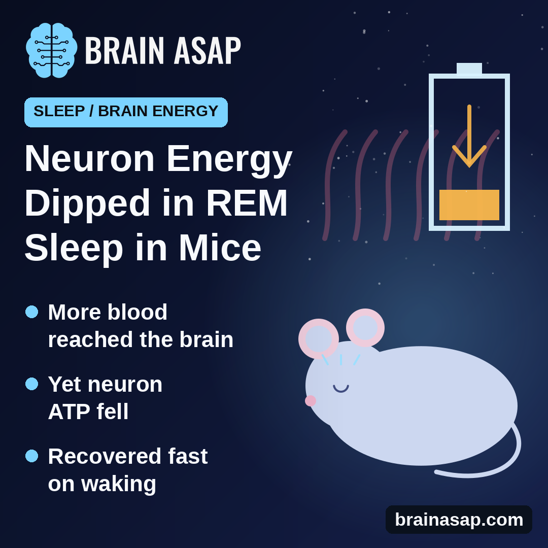
TL;DR: In sleeping mice, blood volume in the brain rose sharply during REM sleep, yet the energy molecule ATP inside neurons dropped. It bounced back as soon as the mice woke, suggesting REM sleep spends energy faster than blood supply alone can explain.
Key Findings
- Imaged through the intact skull: Blood volume, support-cell fuel and neuron energy in naturally sleeping mice.
- Blood surged into REM: Starting about 50 seconds before REM, from the back of the brain forward.
- Support cells stocked up: Fuel (pyruvate) in astrocytes rose during REM.
- Neurons ran lower: ATP fell during REM, most in rear brain areas.
- Fast recovery: ATP returned to normal right after waking.
- Small study: 3 mice per sensor.
Source: Communications Biology (2026) | Takahashi et al.
REM sleep, the stage linked to vivid dreams, is not a restful time for the brain. Neurons fire in wake-like patterns, and blood rushes in.
More blood should mean more fuel. So a team at Tohoku University in Japan expected neurons to be well stocked with energy during REM. In mice, the opposite happened.
Watching Brain Fuel Through the Skull
Brain cells run on ATP, a molecule that stores usable energy. Much of the raw fuel arrives as glucose in the blood. Some goes straight into neurons, and some is processed first by astrocytes, star-shaped support cells that can pass fuel on to neurons.
The researchers bred mice whose astrocytes or neurons carried glowing sensors for pyruvate, an early fuel product, or for ATP. Instead of cutting a window in the skull, which can disturb blood vessels, they coated the intact skull with clear resin and filmed the whole brain surface through it. Brain-wave and muscle recordings showed when each mouse was in non-REM sleep, REM sleep or awake.
Blood Rushed In Before REM Began
During non-REM sleep, blood volume tracked brain activity closely. Changes in theta waves predicted blood changes 4 to 5 seconds later.
Around REM, the pattern changed:
- Early surge: Blood volume began climbing about 50 seconds before REM officially started.
- Back to front: The rise began in rear areas and spread forward.
- No energy change yet: Before REM, pyruvate and ATP held steady.
Neurons Ran Low Anyway
Once REM began, the fuel signals split apart. Pyruvate in astrocytes went up. ATP in neurons went down, most strongly in the retrosplenial cortex, a memory-linked area that works closely with the hippocampus during REM, and in the visual cortex.
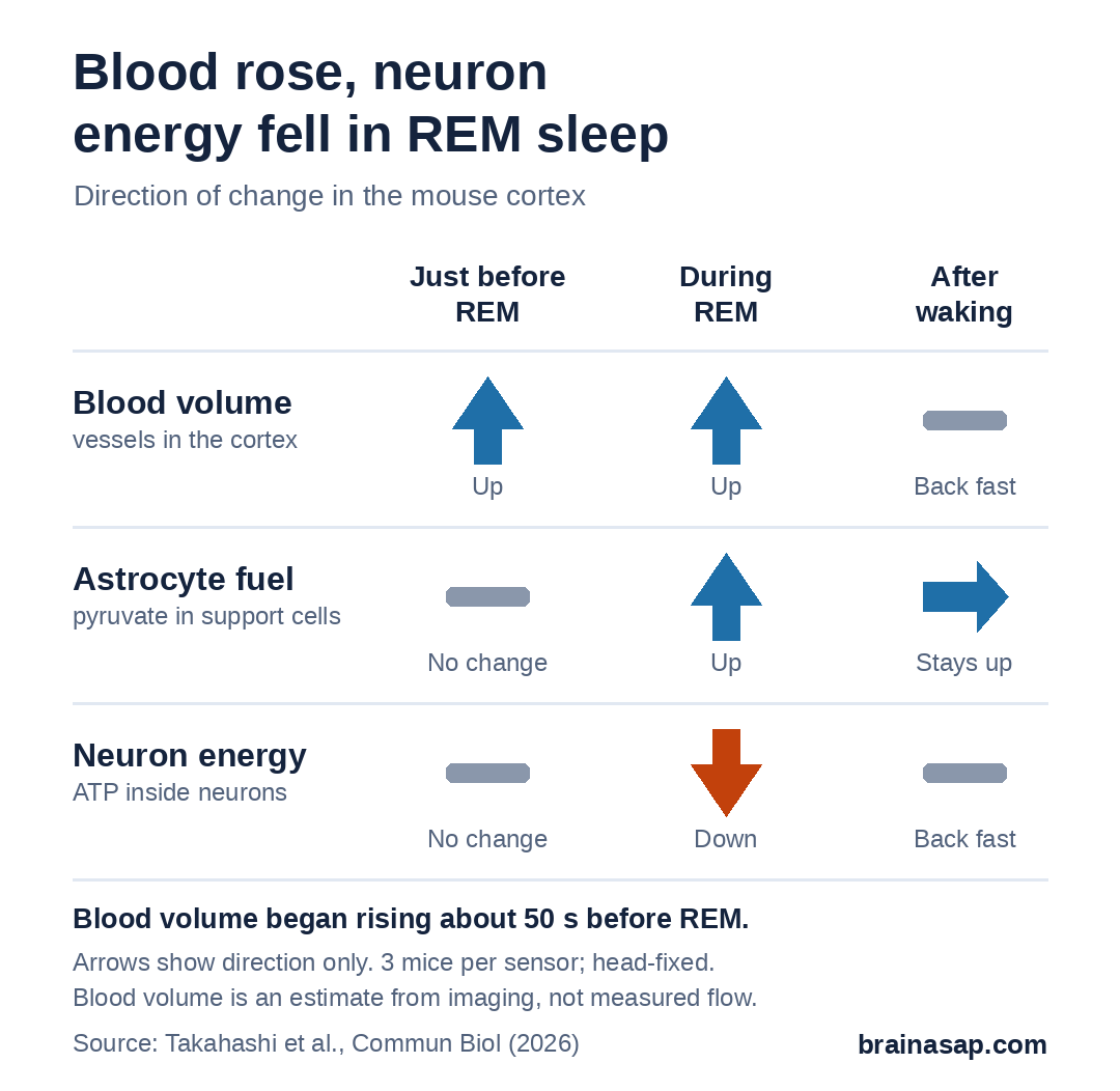
On waking, ATP snapped back to normal almost immediately, while astrocyte pyruvate stayed high for a while. When the team widened blood vessels with a drug outside of sleep recordings, pyruvate and ATP both rose, so extra blood by itself does raise neuron energy. Something specific to REM was pulling ATP down.
Three Possible Explanations
The authors lay out three possibilities that could act together:
- Higher spending: REM may use energy for memory-related rewiring, not just firing.
- Slower handoff: Astrocytes may pass less fuel to neurons, which would explain pyruvate piling up in astrocytes.
- Different engine: Cells may shift away from oxygen-based energy production during REM. Human brain scans have shown more glucose use without more oxygen use in REM.
Firing rates alone probably do not explain it. Other studies show REM firing rises less than 1.3-fold in these areas, and falls in some.
What This Mouse Study Can and Cannot Say
- Mice, not people.
- Few animals: 3 mice per sensor, 15 male mice in total.
- Head-fixed: Mice slept with their heads held under the microscope, for sessions under 4 hours.
- Indirect signals: Blood volume came from imaging, not a direct flow measurement.
- Surface only: Wide-field imaging sees the cortex, not deeper areas.
- Not about dreams: The study did not test dreaming or memory.
A Warning for Reading Brain Scans
The most practical point may be for brain imaging. Functional MRI tracks blood-related signals and is often read as a stand-in for how hard brain cells are working. The authors note that in REM sleep, blood and neuron energy moved in opposite directions, so blood signals may not always reflect neurons’ energy state.
The open question is what REM is spending that energy on, and whether the same dip happens in the human brain.
Citation: DOI: 10.1038/s42003-026-10646-6. Takahashi Y, Ikoma Y, Matsui K. Energy paradox in REM sleep: balancing supply and consumption in brain metabolism. Commun Biol. 2026;9:979.
Study Design: Transcranial wide-field fluorescence imaging with FRET sensors and ECoG/EMG in head-fixed, naturally sleeping transgenic mice.
Sample Size: 15 male mice in total; 3 per sensor experiment (26 REM episodes for pyruvate/ATP maps).
Key Statistic: Blood volume rose from about 50 s before REM; neuronal ATP fell during REM and recovered on waking.
Caveat: Mouse physiology study; small numbers; indirect imaging signals; mechanisms not tested directly.