TL;DR: A 2026 Nature Neuroscience study found that multiple sclerosis lesions rich in lipid-filled foamy microglia had a molecular profile tied to faster disability progression, cerebrospinal-fluid oxylipins, and a druggable lipid enzyme called MAGL.
Key Findings
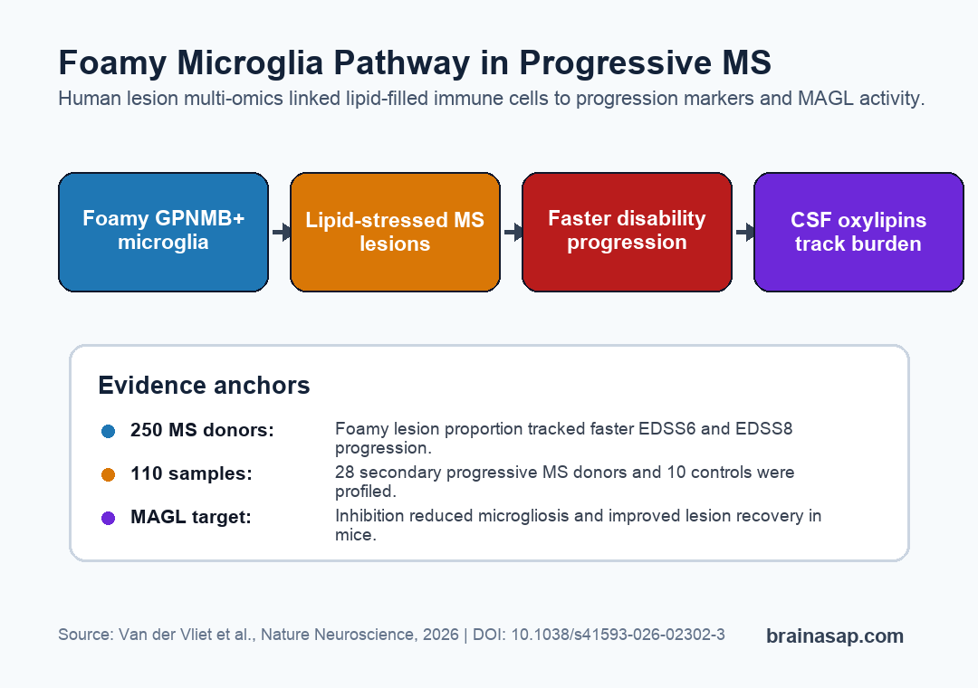
- 250 MS donors: Across Netherlands Brain Bank records, a higher proportion of lesions with foamy microglia correlated with faster progression to severe disability scores.
- 110 white-matter samples: The multi-omics cohort included 28 secondary progressive MS donors and 10 controls, with 58 classified lesion samples.
- Foamy lesions carried a lipid-stress signature: Lesions with foamy microglia were enriched for cholesterol esters, bismonoacylglycerolphosphates, and oxylipins rather than a classic inflammatory cytokine profile.
- MAGL emerged as a target: The lipid enzyme monoacylglycerol lipase was more active in mixed active/inactive lesions with foamy microglia, and MAGL inhibition improved lesion recovery in a mouse demyelination model.
- Cerebrospinal-fluid oxylipins tracked the lesion phenotype: Several lipid mediators in spinal fluid, including 12-HHTrE, PGD2, and 15-HETE, correlated with the proportion of foamy lesions.
Progressive multiple sclerosis lesions can keep expanding even when the early relapsing inflammatory phase is less obvious. Many current therapies suppress relapses better than they stop long-term disability accumulation.
Van der Vliet et al. examined a specific lesion feature that pathologists can see under the microscope: foamy microglia/macrophages, immune cells filled with lipid droplets.
The central question was whether those cells are debris-filled bystanders or part of a lesion state linked to progression.
Foamy Microglia Marked Faster MS Progression
The clinical progression link came first. Researchers connected white-matter pathology from 250 people with multiple sclerosis in the Netherlands Brain Bank to disease-course measures.
Those measures included time to Expanded Disability Status Scale scores of 6 and 8, two severe disability milestones often shortened to EDSS6 and EDSS8.
Donors with a higher proportion of active or mixed active/inactive lesions containing foamy microglia tended to reach disability milestones faster. By contrast, active and mixed active/inactive lesions without the foamy morphology did not show the same progression link, while remyelinated lesions tracked with slower progression.
The comparison narrows the interpretation. The association was not simply “more lesions means worse MS.” The specific foamy lesion phenotype carried the stronger progression marker.
- Foamy lesion load: Associated with faster progression to EDSS6 and EDSS8.
- Nonfoamy active lesions: Did not show the same disease-course relationship.
- Remyelinated lesions: Associated with a slower progression profile.
Multi-Omics Pointed to Lipid Stress, Not Classic Inflammation
The mechanistic work used 110 white-matter samples from 28 secondary progressive MS donors and 10 control donors. The team classified lesions histologically, then profiled them with lipidomics, RNA sequencing, proteomics, chemical proteomics, and targeted histology.
Foamy lesions showed the expected signs of lipid accumulation. Cholesterol esters and triacylglycerols were higher, matching the lipid-filled appearance of the cells.
The broader molecular profile included increased oxylipins, bismonoacylglycerolphosphates, lysosomal stress markers, phagocytosis markers, and antigen-presentation machinery.
The profile did not look like a simple acute inflammatory flare. Several classic cytokines, including TNF, IL6, IFN-gamma, and IL1-beta, were not different between foamy and nonfoamy lesions in the cytokine screen.
The study instead points to a chronic, lipid-processing lesion state.
- Lipid accumulation: Cholesterol esters and neutral lipid staining were higher in foamy lesions.
- Oxylipin enrichment: Oxidized lipid mediators were increased in lesions with foamy microglia.
- Lysosomal stress: Transcriptomic and proteomic signals pointed toward overloaded lipid handling and cell-debris processing.
- B-cell context: Foamy-lesion molecular profiles also included IgG1 and plasma-cell-related signals.
GPNMB Identified a Distinct Microglial State
The study linked foamy cells to a GPNMB-positive microglial state. GPNMB is a glycoprotein often discussed in lysosomal dysfunction and lipid-loaded macrophage biology, so its appearance fits the paper’s lipid-stress theme.
Single-nucleus reference data and bulk profiling pointed to a microglial state the authors call Micro_D. This state was enriched for genes such as GPNMB, APOC1, PLIN2, TREM2, ALOX15B, and CHIT1.
Those genes sit near lipid handling, lysosomal biology, and chronic immune activation rather than a short-lived cytokine burst.
Histology strengthened the point. In mixed active/inactive lesions, the proportion of GPNMB-positive microglia was higher in foamy rims than in nonfoamy rims.
That result gives the microscopy finding a molecular label readers can track.
MAGL Became the Druggable Part of the Pathway
The most actionable part of the paper is the monoacylglycerol lipase (MAGL) finding. MAGL breaks down the endocannabinoid 2-arachidonoylglycerol, or 2-AG, into arachidonic acid, a precursor for many oxylipins.
Activity-based protein profiling found 97 active hydrolases in MS lesions. MAGL activity was generally lower in lesions than in control or normal-appearing white matter, reflecting tissue damage, but it was restored and about fourfold higher in mixed active/inactive lesions with foamy microglia than in nonfoamy mixed lesions.
The team then tested MAGL inhibition in a mouse spinal-cord demyelination model. Treatment with the inhibitor MAGLi-432 began 2 days after lesion induction, which makes the result more relevant to repair than to preventing the lesion from forming.
By 14 days, treated animals showed reduced microgliosis and lipid accumulation, better myelin-debris clearance, and improved lesion recovery.
- Human lesion clue: MAGL activity localized to foamy microglia and astrocytes at lesion rims.
- Biochemical route: MAGL can feed arachidonic-acid-derived oxylipin production.
- Mouse validation: MAGL inhibition improved recovery after demyelination was already triggered.
Cerebrospinal-Fluid Oxylipins Could Mark Foamy Lesions
The biomarker angle is more exploratory but still useful. Because oxylipins are secreted lipid mediators, researchers tested whether cerebrospinal-fluid levels reflected the foamy-lesion burden.
Several cerebrospinal-fluid oxylipins correlated with the proportion of foamy lesions. The study highlights 12-HHTrE, PGD2, and 15-HETE, all of which also aligned with the integrated molecular factor tied to foamy microglia.
A spinal-fluid oxylipin test is not ready for clinic. The tissue study was postmortem, the cerebrospinal-fluid analysis was correlational, and age could complicate lipid measurements.
The practical next step is narrower: measure whether lipid mediators can identify patients whose lesions are stuck in a damaging, foamy microglial state.
Lipid-Loaded Lesions Give Progressive MS a Repair Target
Foamy microglia are not the only possible drivers of progressive MS. This study shows a lesion subtype where lipid handling, immune-cell debris processing, B-cell activity, and oxylipin production converge.
The convergence is important because progressive MS needs treatment strategies that promote lesion resolution, not only relapse suppression. A pathway involving MAGL, 2-AG, arachidonic acid, and oxylipins is specific enough to test in models and eventually in patient-stratified trials.
The main caveat is timing. Human postmortem tissue captures end-stage lesion biology, not the full sequence from lesion initiation to expansion.
The mouse experiment supports a repair role for MAGL inhibition, but it does not prove the same effect in human progressive MS.
The study gives progressive MS a clearer molecular model: some chronic lesions may fail to resolve because lipid-filled microglia become locked into a metabolic immune state.
If that state can be measured and altered, lesion repair becomes a more concrete therapeutic target.
Citation: DOI: 10.1038/s41593-026-02302-3. Van der Vliet et al. Foamy microglia link oxylipins to disease progression in multiple sclerosis. Nature Neuroscience. 2026;29:1585-1598.
Study Design: Human postmortem lesion multi-omics study with supporting mouse demyelination-model validation.
Sample Size: 250 MS donors for pathology-course associations; 28 secondary progressive MS donors and 10 controls for the 110-sample molecular cohort.
Key Statistic: The 250-donor analysis linked higher foamy-lesion proportion with faster time to EDSS6 and EDSS8, while MAGL inhibition improved recovery in a mouse lesion model.
Caveat: Postmortem human tissue and correlational cerebrospinal-fluid measures cannot establish the full temporal sequence of foamy microglia, oxylipins, and disability progression.