TL;DR: A 2026 preprint UK Biobank neuroimaging study in medRxiv found that the comorbidity group showed widespread lower cortical volume, subcortical differences, and white matter microstructure alterations, while several alterations appeared unique to the chronic pain-depression comorbidity group.
Key Findings
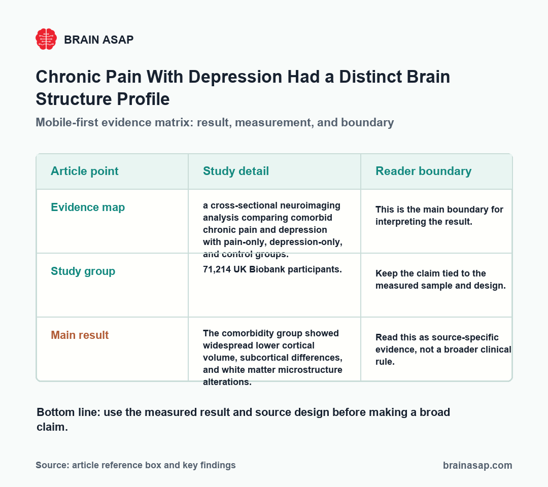
- Evidence map: a cross-sectional neuroimaging analysis comparing comorbid chronic pain and depression with pain-only, depression-only, and control groups.
- Study group: 71,214 UK Biobank participants.
- Main result: The comorbidity group showed widespread lower cortical volume, subcortical differences, and white matter microstructure alterations.
- Second result: Several alterations appeared unique to the chronic pain-depression comorbidity group.
- Caution: Cross-sectional imaging cannot determine whether brain differences preceded, followed, or tracked the comorbidity.
Source: medRxiv (2026) | Casey et al.
Chronic pain and depression often occur together, but many imaging studies examine them separately. That can miss the biology of the combined condition.
This preprint used UK Biobank data to compare four groups: comorbid chronic pain and depression, chronic pain only, depression only, and controls.
The useful finding is that comorbidity had its own imaging profile: people with chronic pain and depression showed widespread cortical, subcortical, and white-matter differences. The finding is not just pain-only plus depression-only pasted together.
UK Biobank MRI Compared Pain, Depression, and Comorbidity
Design: a cross-sectional neuroimaging analysis comparing comorbid chronic pain and depression with pain-only, depression-only, and control groups. Study group: 71,214 UK Biobank participants.
UK Biobank allowed comparison across pain-only, depression-only, comorbid pain-depression, and control groups. This comparison structure is why the comorbidity result is clinically relevant.
- Cortex: The comorbidity group showed regional surface area and thickness differences.
- Subcortex: Thalamic, hippocampal, and left accumbens volumes were lower.
- White matter: Fractional anisotropy and mean diffusivity showed widespread differences.
- Comparison groups: Pain-only and depression-only groups had partly distinct patterns.
The four-group comparison matters because pain and depression overlap clinically but do not have to share one brain profile.
A pain-only group can show sensory and pain-network features, while a depression-only group can show mood, reward, and cognitive-control features.
The comorbidity group tests whether the overlap has its own pattern.
The MRI measures covered several tissue and region classes rather than one headline brain area. The finding is less about a single diagnostic spot and more about a distributed structural profile.
- Cortical measures: surface area and thickness differences can reflect large-scale cortical organization.
- Subcortical volumes: thalamus, hippocampus, and accumbens findings connect pain, memory, mood, and reward systems.
- White matter: fractional anisotropy and mean diffusivity describe microstructure in communication pathways.
- Comorbidity question: the key issue is whether chronic pain with depression should be modeled separately from either condition alone.
The useful interpretation is not that MRI can diagnose pain-depression comorbidity. The result supports a research model in which the combined condition deserves its own longitudinal tests.
Comorbid Pain-Depression Showed Widespread Brain Differences
The main MRI result was broad: lower cortical volume, subcortical differences, and white matter microstructure alterations in the comorbidity group. This overlap may deserve its own imaging model.
Some alterations appeared unique to the chronic pain-depression group. That is more informative than simply saying pain and depression both affect the brain.
Thalamus, Hippocampus, and Accumbens Volumes Were Lower
Structural MRI shows anatomy and tissue-level proxies. It cannot reveal whether the brain differences caused symptoms, followed symptoms, or changed alongside long-term disease burden.
The restrained interpretation is that chronic pain with depression had a distinct cross-sectional brain profile. Longitudinal imaging is needed before direction of effect can be inferred.
Subcortical findings in the thalamus, hippocampus, and accumbens are plausible because those regions sit near pain, mood, memory, and reward systems.
The comorbidity framing also helps prevent a common overread. A large imaging sample can detect statistically reliable differences, but reliable does not automatically mean diagnostic or decisive for clinical care for one patient.
The next test is longitudinal. Researchers need to know whether the structural profile predicts persistent pain, worsening depression, functional recovery, or treatment response.
- Prediction: do baseline MRI features forecast later symptom course?
- Specificity: do the findings remain when pain type and depression severity are separated?
- Treatment response: do brain differences change when pain or depression improves?
Until those questions are tested, the study is strongest as a map of comorbidity biology, not as a clinical imaging test.
Cross-Sectional MRI Cannot Resolve Direction of Effect
Main limitation: cross-sectional imaging cannot determine whether brain differences preceded, followed, or tracked the comorbidity.
- Cross-sectional: Brain differences cannot establish timing or causality.
- UK Biobank: Participants may not represent all clinical pain populations.
- Symptoms: Pain type and depression severity can vary widely.
- Effect sizes: Large samples can detect small differences.
The main limitation is cross-sectional design. Brain structure and clinical burden were measured at one broad time point, so causality remains unresolved.
The clinical use is subgroup definition. If future studies confirm the comorbidity profile, trials could stratify patients with both chronic pain and depression instead of mixing them into pain-only or depression-only categories.
That would make treatment studies easier to interpret. A therapy that helps pain without improving mood, or improves mood without changing pain, may look different in the comorbidity group than in either condition alone.
Comorbid Pain and Depression May Need Separate Imaging Models
The strongest use of this result is subgroup clarity.
- Best use: Treat chronic pain with depression as a comorbidity group that may not reduce to either condition alone.
- Do not overread: Do not claim MRI can diagnose the comorbidity or establish cause from this design.
- Next question: Use longitudinal MRI to test whether brain differences predict symptom persistence, recovery, or treatment response.
That is a cleaner message than a generic brain-change story: the overlap itself may be biologically meaningful.
Citation: DOI: 10.64898/2026.04.02.26350033. Casey et al. Structural brain characteristics of current co-occurring chronic pain and depression. medRxiv. 2026.
Study Design: A cross-sectional neuroimaging analysis comparing comorbid chronic pain and depression with pain-only, depression-only, and control groups.
Sample Size: 71,214 UK Biobank participants.
Key Statistic: The comorbidity group showed widespread lower cortical volume, subcortical differences, and white matter microstructure alterations.
Caveat: Cross-sectional MRI cannot show whether brain differences preceded, followed, or tracked pain-depression comorbidity.