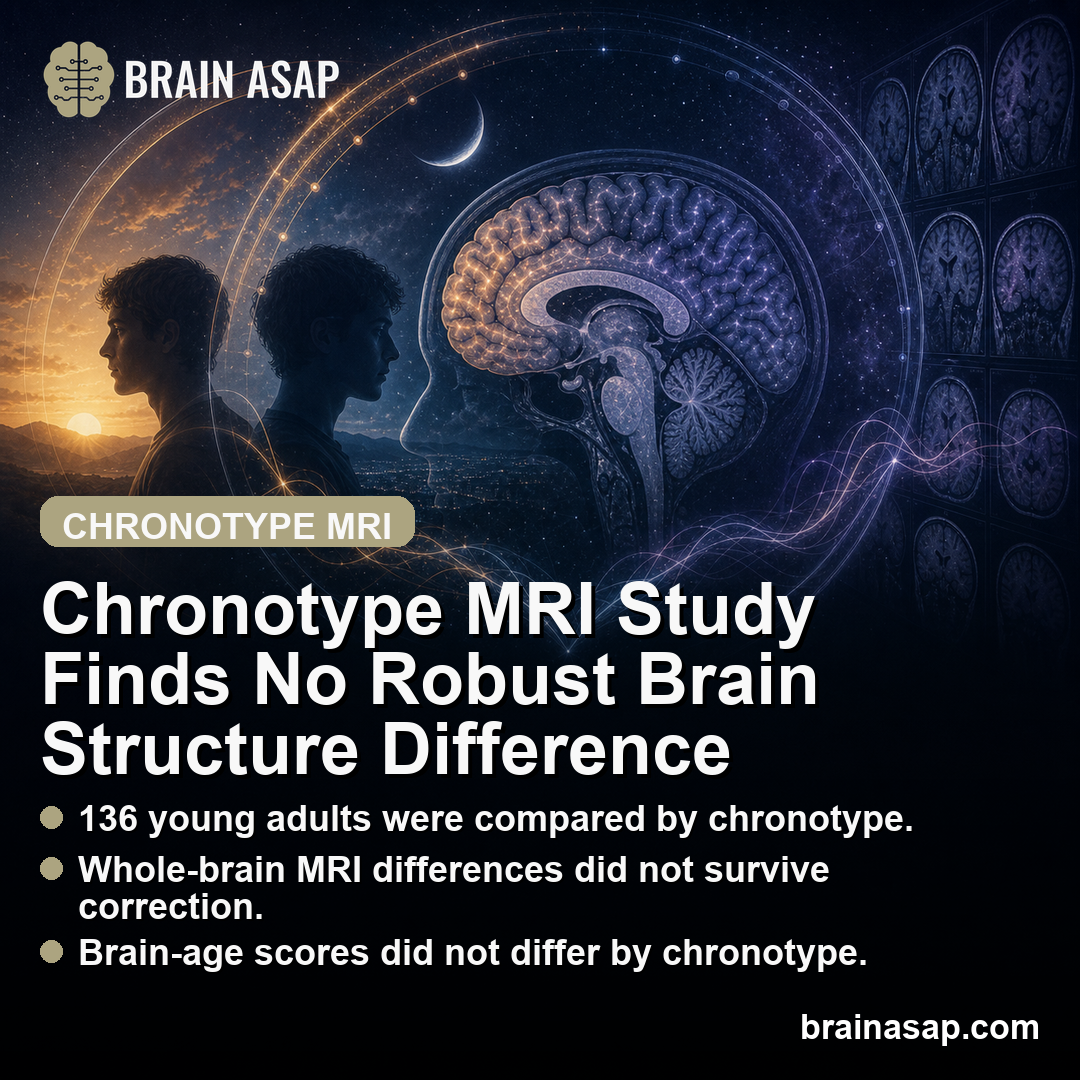
TL;DR: A 2026 MRI study in Brain Imaging and Behavior of 136 healthy young adults found no robust whole-brain gray matter, white matter, cortical thickness, or brain-age differences between early and late chronotypes.
Key Findings
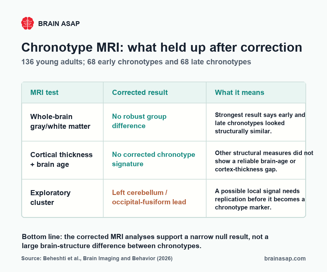
- 136 young adults: The study compared 68 early chronotypes with 68 late chronotypes.
- No robust whole-brain VBM differences: Voxel-based gray and white matter analyses did not survive family-wise error correction.
- Exploratory gray matter cluster: Late chronotypes showed lower gray matter in a left cerebellum/occipital-fusiform cluster under a more exploratory threshold.
- Cortical thickness was not significant after correction: Nominal differences in cingulate, occipital, and temporal regions did not survive false-discovery-rate correction.
- No brain-age gap: Brain-PAD values did not differ significantly between chronotype groups.
Source: Brain Imaging and Behavior (2026) | Beheshti et al.
Chronotype MRI Tested Morningness and Eveningness in Young Adults
Chronotype describes a person’s preferred timing for sleep and activity. Early chronotypes tend to feel and function better earlier in the day, while late chronotypes tend to prefer later sleep and wake times.
Chronotype is linked to circadian biology, sleep timing, mental health, school and work schedules, and lifestyle patterns.
Researchers have therefore asked whether early and late chronotypes also show measurable brain-structure differences.
This 2026 MRI study tested that idea in healthy young adults. It compared brain anatomy across early and late chronotype groups using multiple structural MRI approaches, including voxel-based morphometry, cortical thickness analysis, and brain-age prediction.
The corrected result was cautious: the study did not find robust whole-brain structural differences between groups. The evidence pushes back against easy claims that being a morning person or evening person must leave a large anatomical signature in the young adult brain.
Chronotype is still real when structural MRI looks similar. Sleep timing can shape attention, mood, academic performance, and social jet lag.
The MRI test here was narrower: whether those timing preferences corresponded to reliable anatomical differences in a healthy young sample.
Early and Late Chronotype Groups Were Matched in Size
The sample included 136 healthy young adults, split evenly into 68 early chronotypes and 68 late chronotypes. The researchers used MRI to compare several types of structural brain measures.
The researchers used three structural MRI measures:
- Voxel-based morphometry (VBM): tested gray matter volume and white matter volume across the brain.
- Cortical thickness: examined the thickness of the cerebral cortex in predefined regions.
- Brain-PAD: estimated whether a participant’s brain looked older or younger than expected for chronological age.
Using several MRI measures helped prevent overreliance on one method.
A finding that appears across gray matter volume, cortical thickness, and brain-age models would be more convincing than a single exploratory cluster.
In this study, that converging pattern did not appear.
The balanced group size also reduces one common source of ambiguity.
With 68 people in each chronotype group, the comparison was not driven by a heavily uneven sample.
The study still had limits, but the basic early-versus-late contrast was straightforward.
Whole-Brain VBM Found No Corrected Gray or White Matter Difference
The voxel-wise gray matter and white matter analyses found no group differences that survived family-wise error correction at p < 0.05. In MRI statistics, that correction guards against false positives because thousands of brain locations are tested at once.
Without correction, a brain map can produce scattered apparent differences simply because so many comparisons are being made. Family-wise error correction sets a stricter bar, reducing the risk that the reported finding is a false positive.
The corrected result was straightforward: in this cohort of young adults, early and late chronotypes did not show a robust whole-brain gray matter or white matter volume difference.
The strongest claim should stay narrow: chronotype may shape sleep timing and daily function without leaving a large structural MRI signature in healthy young adults.
Instead of assuming chronotype has a large structural footprint, researchers may need to look at sleep behavior, circadian physiology, functional connectivity, or stress exposure.
Exploratory MRI Suggested a Left Cerebellum Occipital-Fusiform Cluster
The exploratory analysis did identify one gray matter cluster where late chronotypes had lower values than early chronotypes. The cluster included left cerebellar and occipital-fusiform regions.
The reported cluster size was 4,097 voxels, with cluster-level false-discovery-rate q = 0.005 and peak T = 4.05. The peak coordinate was MNI -22, -69, -24.
The exploratory cluster can guide future hypotheses, but it should not be treated as a settled chronotype brain marker. It came from a more exploratory threshold rather than the strongest corrected whole-brain result.
The region is also not specific to chronotype.
Cerebellar and occipital-fusiform areas can be involved in visual processing, timing, coordination, and broader cognitive functions.
A structural association there would need replication and behavioral linkage before it could be interpreted mechanistically.
A stronger follow-up would test whether the same cluster appears in an independent cohort and whether it relates to measured sleep timing, alertness, or circadian misalignment. Without that extra evidence, the cluster is best treated as a lead for future research rather than a practical biomarker.
Cortical Thickness and Brain-PAD Did Not Support a Strong Chronotype Signature
Cortical thickness results were also limited. The paper reported nominal differences in bilateral caudal anterior cingulate cortex, right lateral occipital cortex, and left temporal pole, but those findings did not survive false-discovery-rate correction.
Mean cortical thickness was almost identical between groups, at 2.63 +/- 0.07 for early chronotypes and 2.64 +/- 0.05 for late chronotypes. The reported p value was approximately 1, indicating no meaningful group difference in the global measure.
Brain-age analysis told the same cautious story.
Brain-PAD was 0.6 +/- 4.3 years for late chronotypes and 0.4 +/- 5.2 years for early chronotypes.
The group difference was not significant, with t(134) = -1.45 and p = 0.15, and the overall model was not significant (R2 = 0.03, p = 0.32).
Brain-PAD is often used as a rough marker of whether brain structure appears older or younger than expected. Here, it did not suggest that late chronotype young adults had accelerated or delayed structural brain aging.
This null result is relevant because late chronotype is sometimes associated with shorter sleep, later social schedules, and mental health risk.
In this sample, those broader associations did not translate into a detectable brain-age difference. The brain-age model therefore reinforced the main corrected MRI result.
Chronotype Brain Claims Need Corrected Statistics
The interpretation should stay tied to the MRI question. Chronotype may still relate to sleep quality, mood, circadian alignment, cognition, and daily functioning.
This study suggests those differences do not map onto a large, robust structural MRI signature in healthy young adults.
Several limits keep the result focused:
- Young healthy sample: Findings may differ in adolescents, older adults, shift workers, or clinical populations.
- Cross-sectional design: The study cannot show whether chronotype shapes brain structure over time.
- Exploratory cluster: The cerebellum/occipital-fusiform result needs replication.
- Structural MRI only: Functional connectivity, sleep physiology, and circadian hormone measures could show different patterns.
The careful conclusion is that early and late chronotypes looked structurally similar by the strongest MRI tests used here. Chronotype can affect daily life without requiring a large anatomical difference detectable by standard structural MRI.
Sharper follow-up work could test the same question with stronger circadian measurement.
Future studies could combine structural MRI with actigraphy, melatonin timing, sleep diaries, or longitudinal follow-up.
Those methods may reveal whether chronic circadian misalignment, rather than chronotype preference alone, is the stronger brain-health exposure.
Citation: DOI: 10.1007/s11682-026-01153-7. Beheshti et al. Neuroanatomical signatures of chronotype in young adults. Brain Imaging and Behavior. 2026.
Study design: Structural MRI comparison of early and late chronotypes using voxel-based morphometry, cortical thickness analysis, and brain-age prediction.
Sample Size: 136 healthy young adults, including 68 early chronotypes and 68 late chronotypes.
Key Statistic: No whole-brain gray matter, white matter, cortical thickness, or brain-age difference survived corrected statistical thresholds.
Caveat: The localized cerebellum/occipital-fusiform finding was exploratory and needs replication.