TL;DR: A 2026 meta-analysis in Dialogues in Clinical Neuroscience pooled whole-brain functional MRI studies and found that anxious major depressive disorder (anxious MDD, depression with prominent anxiety symptoms) showed different brain-activity patterns from both non-anxious MDD and healthy controls.
Key Findings
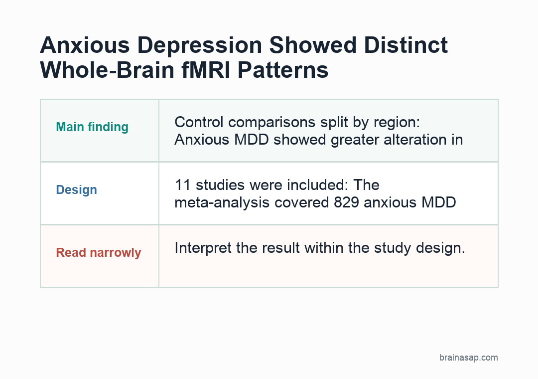
- 11 studies were included: The meta-analysis covered 829 anxious MDD patients, 681 MDD patients, and 865 healthy controls.
- Temporal-lobe activity differed: Anxious MDD showed greater alteration in the left middle temporal gyrus than non-anxious MDD.
- Control comparisons split by region: Anxious MDD showed greater alteration in the anterior commissure and lower alteration in the right middle frontal gyrus versus healthy controls.
- Female subgroup results were separate: Meta-regression linked anxious MDD in women to higher cingulate and thalamic-region activity and lower left rolandic operculum activity.
- No diagnostic scan rule: Researchers pooled published coordinates from whole-brain fMRI studies, so the findings are group-level markers, not a clinical scan readout.
Source: Dialogues in Clinical Neuroscience (2026) | Huang et al.
Major depressive disorder is already heterogeneous. Some patients mainly report low mood and loss of interest, while others also show prominent anxiety, agitation, worry, or panic-like symptoms.
The 2026 review focused on that second pattern: anxious major depressive disorder, abbreviated as anxious MDD. The question was whether this subgroup shows a different functional brain signature when researchers look across whole-brain functional MRI studies.
Whole-Brain fMRI Studies Were Pooled Across 2,375 Participants
Researchers searched for neuroimaging studies that compared anxious MDD, non-anxious MDD, and healthy controls. They included only studies that used whole-brain analysis and reported peak brain-coordinate results.
The whole-brain design is important because a region-of-interest study starts with a preselected brain area. A broader statistical search is better suited for asking whether anxious MDD has a reproducible pattern across studies.
- Anxious MDD group: 829 patients across the included studies.
- MDD comparison group: 681 patients with non-anxious MDD.
- Healthy controls: 865 participants without the target diagnosis.
- Core method: Seed-based d Mapping, a coordinate-based meta-analysis method for pooling reported fMRI peaks.
Functional MRI, or fMRI, refers to imaging methods that estimate brain activity or functional connectivity through blood-oxygen-related signal changes. The meta-analysis did not reprocess raw scans from every participant.
Left Middle Temporal Gyrus Differed From Non-Anxious Depression
The clearest anxious MDD versus MDD result was in the left middle temporal gyrus, a temporal-lobe region involved in language, semantic processing, and social-emotional interpretation. Anxious MDD showed greater functional alteration there than non-anxious MDD.
The reported cluster was centered at MNI coordinates -52, -42, 8, with SDM-Z = 2.046, p = 0.020, and 359 voxels. MNI coordinates are a standard brain-map coordinate system used to describe where an imaging result sits.
The left middle temporal gyrus finding is not an individual diagnostic marker. Across the included whole-brain studies, this region was one place where anxious MDD separated from non-anxious MDD at the group level.
Control Comparisons Pointed to Anterior Commissure and Frontal Cortex
When anxious MDD was compared with healthy controls, two regions moved in opposite directions. The anterior commissure, a white-matter pathway connecting parts of the temporal lobes, showed greater alteration in anxious MDD.
The right middle frontal gyrus showed lower alteration in anxious MDD than controls. The middle frontal gyrus is part of the frontal cortex and is often discussed in relation to cognitive control, attention, and emotion regulation.
- Anterior commissure: Higher alteration in anxious MDD versus controls, with p = 0.004.
- Right middle frontal gyrus: Lower alteration in anxious MDD versus controls, with p = 0.028.
- MDD versus controls: The re-analyzed seven-study comparison did not show a significant pooled difference.
The control comparison is useful because it suggests anxious MDD is not only ordinary MDD plus more worry. The pooled studies pointed to a different mix of temporal, commissural, and frontal-cortex findings.
Female Subgroup Signals Were Found in Cingulate and Thalamic Regions
The researchers also ran meta-regression because anxiety and depression are both more common in women than in men. In the female anxious MDD subgroup, several regions differed from controls.
Higher functional alteration appeared in the left cingulate region and right anterior thalamic projections. The cingulate cortex is involved in emotion, attention, and conflict monitoring, while thalamic projections help relay and coordinate signals across brain networks.
- Left cingulate-region finding: Higher alteration, with p = 0.018.
- Right anterior thalamic projections: Higher alteration, with p = 0.034.
- Left rolandic operculum: Lower alteration, with p = 0.049.
The sex-stratified result should be read carefully. Meta-regression can identify patterns that track with study-level variables, but it is not the same as a prospective study designed from the start to test sex-specific mechanisms in individual patients.
The Findings Are Group-Level Markers, Not Clinical Scan Rules
The main limitation is that the evidence came from published whole-brain fMRI coordinates rather than a shared raw-imaging dataset. Researchers also excluded region-of-interest studies, which kept the analysis methodologically cleaner but left out some potentially relevant imaging work.
Diagnostic boundary: A routine fMRI scan cannot confirm anxious depression.
These findings are better understood as candidate neurobiological markers for future research on depression subtypes.
- Strongest use: Refining hypotheses about anxious MDD as a depression subtype with partly distinct brain-network features.
- Main caution: The pooled results do not prove causality or individual diagnostic accuracy.
- Next step: Larger studies using shared raw imaging, symptom ratings, medication data, and sex-stratified models would be needed before clinical use.
For now, the pooled evidence supports a narrower claim: anxious MDD showed reproducible group-level fMRI differences in temporal, frontal, commissural, and sex-linked regions across the available whole-brain literature.
Citation: DOI: 10.1080/19585969.2026.2612918. Huang et al. Functional brain alterations in anxious depression: Insights from whole-brain fMRI and meta-analysis. Dialogues in Clinical Neuroscience. 2026;28:32-45.
Study Design: Coordinate-based meta-analysis of whole-brain fMRI studies comparing anxious MDD, non-anxious MDD, and healthy controls.
Sample Size: 11 studies; 829 anxious MDD patients, 681 MDD patients, and 865 healthy controls.
Key Statistic: Anxious MDD showed greater left middle temporal gyrus alteration versus MDD (p = 0.020) and lower right middle frontal gyrus alteration versus controls (p = 0.028).
Caveat: The analysis used published coordinate peaks from whole-brain fMRI studies, so the findings are group-level markers rather than individual diagnostic rules.