TL;DR: A 2026 Reviews in the Neurosciences review argues that glycolysis, the cell pathway that turns glucose into usable energy and lactate, is a disease-shaping process across Alzheimer’s disease, Parkinson’s disease, ALS, Huntington’s disease, Wilson disease, and multiple sclerosis rather than a background energy problem.
Key Findings
- Glycolysis changes differed by brain cell type, with neurons, astrocytes, microglia, and oligodendrocytes using glucose and lactate in different ways.
- The review linked altered glucose metabolism to six neurodegenerative disease groups, but emphasized that the same pathway can look underactive, overactive, or compensatory depending on stage and tissue.
- Promising tools include FDG-PET, hyperpolarized carbon-13 magnetic resonance spectroscopy, metabolomics, single-cell/spatial transcriptomics, lactate sensors, and Seahorse metabolic assays.
- Cell-specific targeting was central to the review because the same glycolysis intervention could have different effects in neurons, glia, and immune cells.
- The therapeutic argument is cautious: glycolysis targets such as PFKFB3, lactate shuttling, microglial metabolism, and systemic metabolic modulation need stage-specific safety testing.
Source: Cheng T, Li M, Yang Y, Rao Z, and Yang W, Reviews in the Neurosciences, 2026.
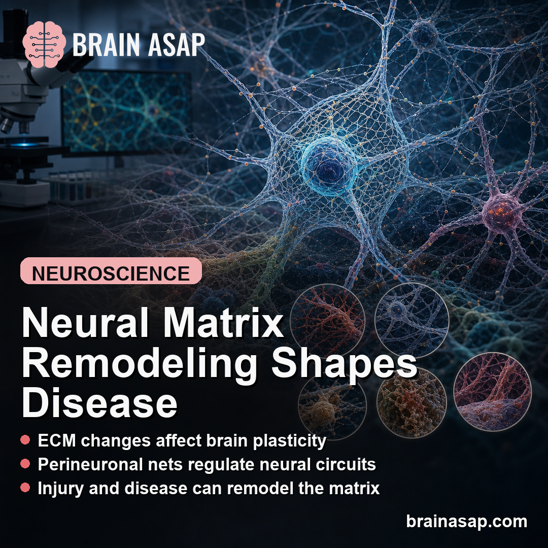
Glycolysis Was Framed as a Disease Axis, Not Just Fuel Use
Glycolysis is the pathway that breaks glucose into pyruvate and can also feed lactate production. In the brain, that pathway sits beside mitochondrial oxidative phosphorylation, the pentose phosphate pathway, antioxidant defense, and biosynthesis.
The review’s main point was not that every neurodegenerative disease has the same glucose problem. It was that glycolytic flux, lactate handling, and glucose-branching decisions can shape vulnerability across cells and disease stages.
Researchers described glycolysis as a hub connecting several processes that usually get discussed separately:
- Energy supply: ATP production can fail when glucose uptake, pyruvate entry into mitochondria, or lactate use breaks down.
- Redox balance: Shifting glucose away from the pentose phosphate pathway can reduce NADPH, a key support for antioxidant defenses.
- Inflammation: Activated microglia and immune cells can move toward a more glycolytic state that supports inflammatory behavior.
- Cell support: Astrocytes and oligodendrocytes can use lactate shuttling to support neurons and axons.
This framing changes how a low-glucose imaging pattern should be read. It can reflect cell-specific remodeling: reduced neuronal energy capacity in one place, compensatory astrocyte glycolysis in another, and inflammatory glycolysis in immune cells or microglia.
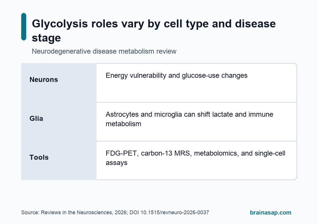
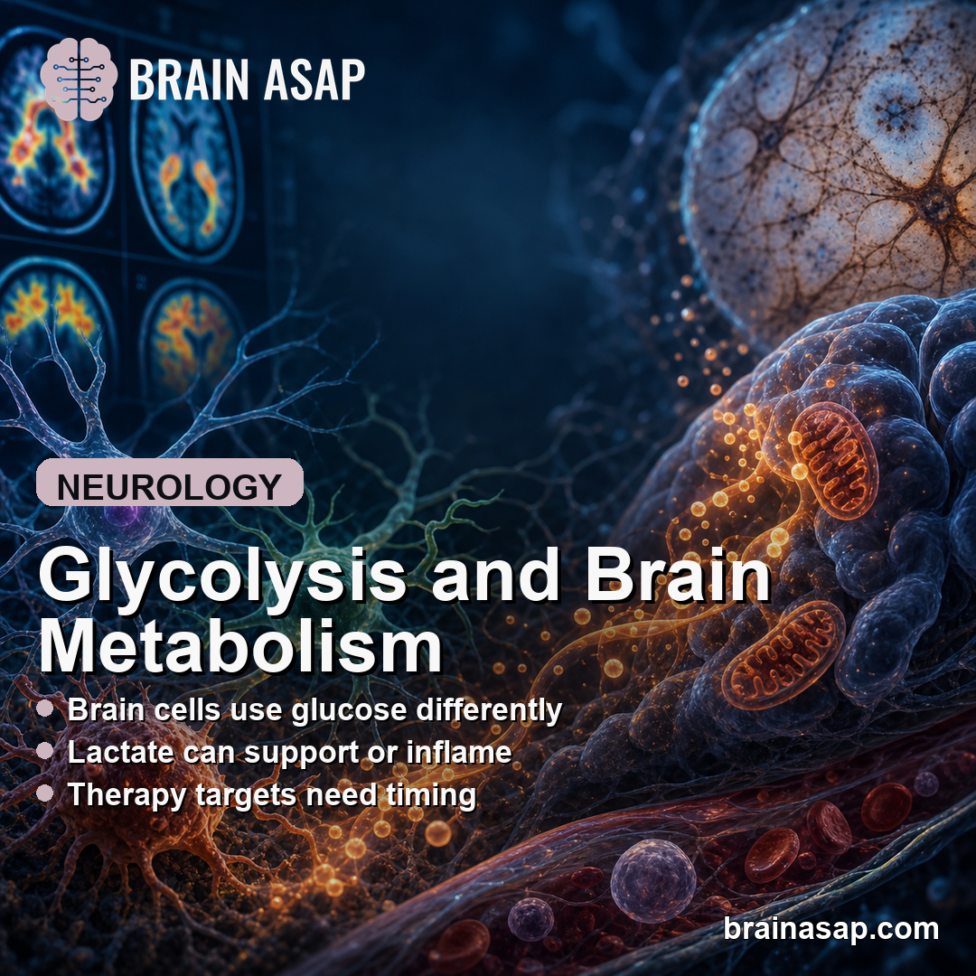
Brain Cells Use Glucose and Lactate Differently
The review separated the brain’s metabolic labor by cell type. Neurons usually rely heavily on mitochondrial oxidative phosphorylation and the pentose phosphate pathway, which helps maintain antioxidant capacity.
Astrocytes are more glycolytic. They can convert glucose into lactate and export that lactate through monocarboxylate transporters.
Neurons can use that lactate during synaptic activity. This is the astrocyte-neuron lactate shuttle, often shortened to ANLS.
Microglia, the brain’s resident immune cells, can shift metabolic gears. Resting microglia depend more on oxidative phosphorylation, but inflammatory activation can push them toward glycolysis, lactate production, and inflammatory gene programs.
Oligodendrocytes, the cells that support myelin, use glycolysis in a spatially organized way. The review noted that glycolytic enzymes and glycolysis-derived metabolites can be enriched in non-compacted myelin regions, where local energy support matters for long-range axons.
Those differences make a single “boost glycolysis” or “block glycolysis” strategy risky. The same intervention can help one cell type while worsening oxidative stress, inflammation, or support-cell failure in another.
Alzheimer’s and Parkinson’s Show Stage-Specific Metabolic Patterns
For Alzheimer’s disease, the review highlighted a familiar clinical pattern: FDG-PET hypometabolism in vulnerable regions such as posterior cingulate, parietal, and temporal cortex can appear early and correlate with cognitive decline.
At the same time, Alzheimer’s-related glycolysis was not described as only low metabolism. The review discussed reduced glycolytic intermediates in cerebrospinal fluid, neuronal PFKFB3 changes that can redirect glucose away from antioxidant support, and reactive astrocyte states that may increase glycolysis or lactate production.
The disease-specific examples split into several practical patterns:
- Alzheimer’s disease: Regional glucose hypometabolism, amyloid/tau-linked metabolic stress, PFKFB3 dysregulation, and disrupted lactate shuttling.
- Parkinson’s disease: Abnormal glucose metabolism in cortical and subcortical patterns, with changes relevant to early disease and cognitive decline.
- Frontotemporal dementia: Frontal-temporal glucose reductions, possible astrocyte autophagy and metabolic failure, and mixed cell-type remodeling.
- ALS: Motor-system and systemic metabolic disturbances, including cases where glycolysis may become compensatory under mitochondrial stress.
The review therefore treated timing as central. Moderate glycolytic support can be protective in one stage, while excessive or misplaced glycolysis can feed oxidative stress, synapse loss, or inflammation in another.
Multiple Sclerosis Added an Immune-Metabolism Loop
Multiple sclerosis was useful because the glycolysis question extends beyond neurons. The review described immune-cell glycolysis as part of a feedback loop involving peripheral immune activation, tissue infiltration, central nervous system injury, and more metabolic reprogramming.
Within MS lesions, disrupted astrocyte-axon support and altered lactate handling may briefly help meet high energy demand during inflammation. Chronic dysregulation can instead worsen axonal stress and degeneration.
Lactate also has regulatory roles. It is not only a fuel molecule. The review discussed lactate as a regulator of acidosis, receptor pathways, transporter activity, and epigenetic processes such as lactylation.
The MS section makes the broader caution concrete: a glycolysis-centered therapy needs to account for disease subtype, active inflammation, chronic injury, cell type, and whether the target tissue is brain, spinal cord, immune cell, or peripheral metabolism.
New Tools Can Separate Uptake, Flux, and Cell Type
The review’s methods section was important because different tools answer different questions. FDG-PET shows regional glucose uptake and is already used in dementia subtyping and progression assessment, but it does not directly show where glucose carbon goes after uptake.
Other methods can get closer to glycolytic flux, cell state, or lactate movement:
- Hyperpolarized carbon-13 MRS: Tracks labeled substrates into metabolites such as lactate, pyruvate, and bicarbonate.
- Metabolomics: Measures pathway intermediates in tissue, blood, cerebrospinal fluid, or cell models.
- Single-cell and spatial transcriptomics: Links glycolysis-related gene programs to cell type and tissue location.
- Lactate sensors: Genetically encoded indicators can track lactate dynamics in living cells or tissue.
- Seahorse assays: Measure oxygen consumption and extracellular acidification in cells, separating mitochondrial respiration from glycolytic activity.
Together, those methods can test whether a metabolic finding is neuronal energy failure, astrocyte compensation, microglial inflammation, oligodendrocyte-axon support failure, or a systemic metabolic change that also appears outside the brain.
Therapy Targets Need Timing and Cell-Type Guardrails
The therapeutic section focused on targets rather than settled treatments. One example was PFKFB3, an enzyme regulator that can shift glucose handling in neurons.
In some Alzheimer’s-related models, PFKFB3 changes may redirect glucose away from antioxidant support and worsen oxidative injury.
Other possible targets included the astrocyte-neuron lactate shuttle, microglial glycolysis and lactylation, systemic metabolic interventions, and nanoparticle delivery approaches meant to reach the brain more precisely.
The review did not treat those as ready clinical answers. It named four major barriers:
- Causality: Many findings are cross-sectional or model-based, so glycolysis may be a cause, a driver, a compensation, or a consequence.
- Biphasic effects: Moderate enhancement may help in one setting, while excessive activation may harm another.
- Brain-periphery integration: Blood cells, muscle, liver, and other tissues may show related metabolic changes.
- Standardization: Imaging, biofluid, and cell-assay metrics still need clearer cross-study standards.
Glycolysis is not just a fuel label in neurodegeneration research. It is a testable metabolic control point.
The safest future work will need longitudinal imaging, cell-type-resolved assays, and stage-specific trials before glycolysis can be treated as a reliable therapeutic target.
Citation: DOI: 10.1515/revneuro-2026-0037. Cheng et al. Glycolysis as a central pathological axis in neurodegenerative diseases. Reviews in the Neurosciences. 2026.
Study Design: Narrative review of glycolysis, lactate handling, brain-cell metabolism, research methods, and therapeutic targets across neurodegenerative diseases.
Sample/Model: Human imaging and biomarker literature, cell and animal models, and disease-specific evidence for Alzheimer’s disease, Parkinson’s disease, ALS, Wilson disease, Huntington’s disease, and multiple sclerosis.
Key Statistic: Not a pooled-effect review; the central evidence is cross-disease convergence around glucose uptake, glycolytic flux, lactate metabolism, and cell-type metabolic reprogramming.
Caveat: Much of the causal evidence remains model-based, and glycolysis can be protective, compensatory, or harmful depending on disease stage and cell type.