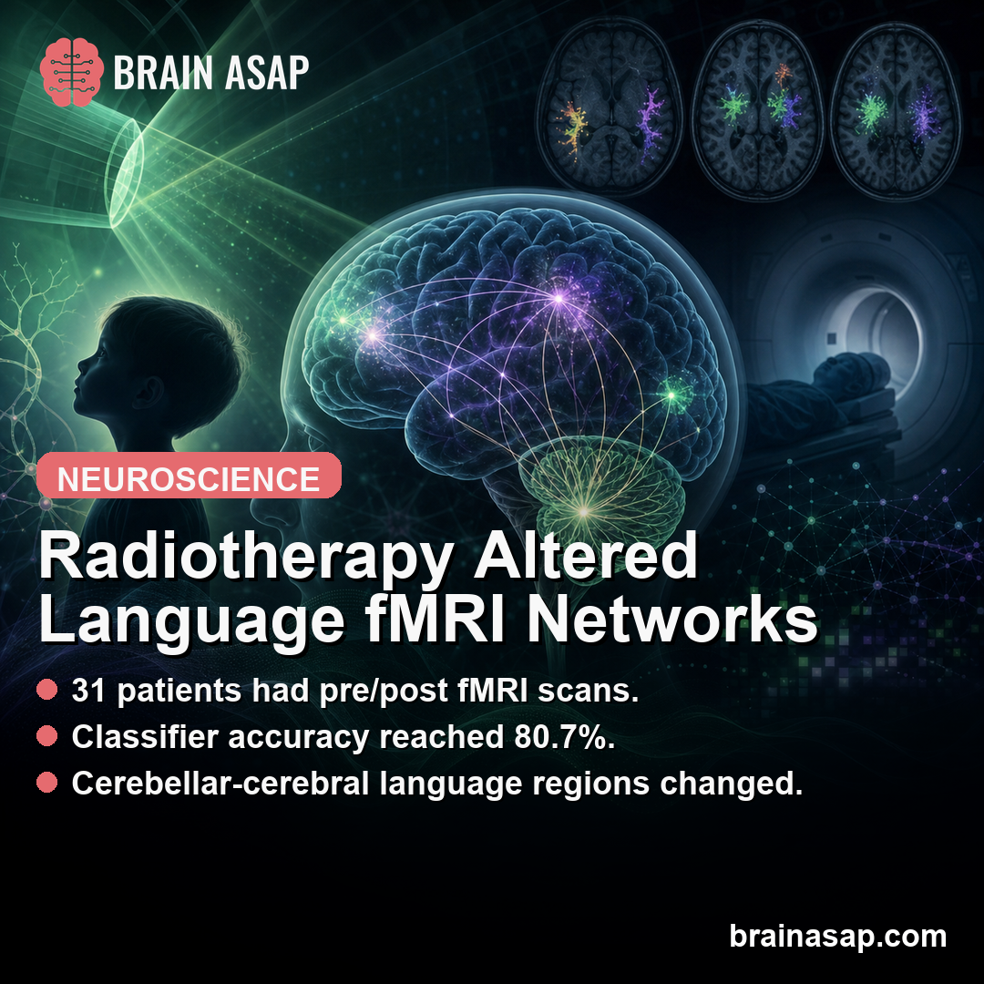
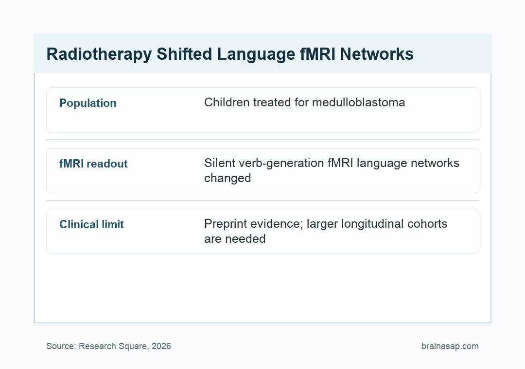
TL;DR: A 2026 Research Square preprint used silent verb-generation functional MRI (fMRI), brain imaging during covert word production, and found early cerebellar-cerebral language-network changes after radiotherapy in 31 children and adolescents treated for medulloblastoma.
Key Findings
- 31 pediatric patients: Children and adolescents with medulloblastoma completed silent verb-generation fMRI before radiotherapy and again within 6 weeks after treatment.
- 80.7% classification accuracy: A nested feature-selection and support vector machine framework distinguished pre- from post-radiotherapy scans.
- Permutation-tested result: The classifier exceeded the label-swap null distribution, with one-sided p = 0.0149.
- Reduced task activity: Post-treatment scans showed reduced activity in bilateral cerebellar hemispheres, left inferior frontal gyrus, left insula, left putamen, anterior cingulate, and left middle temporal gyrus.
- Parietal compensation possibility: Researchers reported increased engagement of the left supramarginal gyrus and left inferior parietal lobule.
Source: Research Square preprint (2026) | da Rocha et al.
Medulloblastoma is a malignant pediatric brain tumor, and radiotherapy can be lifesaving. Treatment can also affect developing brain networks that support language, attention, executive function, and school-age cognition.
The da Rocha preprint examined early functional change rather than waiting for long-term cognitive outcomes. Researchers asked whether a language task during fMRI can detect brain-network reorganization soon after radiotherapy.
Silent Verb-Generation fMRI Tested Language Network Function
The task was silent verb generation. During scanning, patients saw or heard noun cues and covertly generated related verbs, allowing researchers to probe language production without requiring spoken responses in the scanner.
That design fits pediatric neuro-oncology. Overt speech can create motion artifacts, and children recovering from tumor treatment can vary in stamina, speech output, or comfort during scanning.
Silent tasks also reduce the mismatch between the scanner environment and the patient’s ability to cooperate. The child can perform the language operation internally while the imaging signal captures task-related network engagement.
Functional MRI measures blood-oxygen-level-dependent (BOLD) activity, an indirect marker of task-related neural engagement.
In this study, the goal was not to diagnose a language disorder at bedside, but to detect whether treatment changed the activation pattern supporting covert speech.
Researchers scanned 31 children and adolescents enrolled on SJMB12 before radiotherapy and within 6 weeks after completing radiotherapy. The average age was 14.1 years, and 18 participants were male.
The short follow-up window is useful because it captures early treatment-linked functional change. Later cognitive outcomes can include recovery, compensation, tumor effects, schooling disruption, fatigue, and additional therapy exposures.
Machine Learning Distinguished Pre- and Post-Radiotherapy Scans
The analysis used a two-stage nested feature-selection framework with a linear support vector machine.
In plain terms, the model searched for activation features that best separated scans collected before radiotherapy from scans collected afterward.
The classifier reached 80.7% leave-one-subject-out accuracy. Researchers then compared that result against a paired pre-to-post label-swap permutation test, which produced a one-sided p value of 0.0149.
- Feature selection: The workflow selected informative imaging features inside cross-validation.
- Subject-level testing: Leave-one-subject-out validation tested whether the model generalized across participants.
- Permutation benchmark: Label swapping tested whether the pre/post separation exceeded chance structure.
The classifier is not ready for individual clinical decision-making. The imaging pattern contained enough treatment-related information to separate the two scan timepoints better than expected by chance.
That distinction is important. A group-level pre/post classifier can reveal treatment-related network structure without proving that a single child’s scan can predict future language or school performance.
The model therefore functions more like a research probe than a clinical test.
It shows that the pre/post difference was organized enough to classify, not that the algorithm should guide an individual child’s care.
Radiotherapy Was Linked to Reduced Cerebellar and Left Frontal Activity
The post-treatment pattern included reduced task-related activity in bilateral cerebellar hemispheres, including Crus I/II and lobules VI-VIII.
These cerebellar regions are increasingly recognized as part of cognitive and language networks, not only motor coordination.
Researchers also reported reduced activity in left inferior frontal gyrus, left insula, left putamen, supragenual anterior cingulate, and left middle temporal gyrus.
Those areas connect with speech planning, language selection, cognitive control, and verbal processing.
The anatomical pattern is clinically plausible because medulloblastoma treatment often involves posterior fossa and cerebellar vulnerability.
A language-network task can therefore reveal changes that are not limited to the tumor bed or a single anatomical region.
Cerebellar involvement also fits modern language neuroscience. The cerebellum contributes to timing, prediction, error correction, and coordination of cognitive operations, including speech and language tasks.
That broader cerebellar role explains why posterior fossa treatment can have cognitive consequences even when the task seems language-specific.
Language production depends on distributed timing and control systems, not only classic cortical language areas.
For families, this distinction can be hard to see from standard scans alone.
A structural MRI can show tumor control or expected treatment change, while task fMRI can show how the child is recruiting networks during a cognitive operation.
Left Parietal Recruitment May Reflect Working-Memory Compensation
Not every post-treatment change was reduced activity. The analysis also found increased engagement of the left supramarginal gyrus and left inferior parietal lobule.
Researchers interpreted this as possible compensatory recruitment of dorsal parietal phonological working-memory regions. Phonological working memory helps hold speech sounds and word forms online while a person manipulates language internally.
That interpretation fits the task. Silent verb generation requires a child to receive a noun cue, search for a related action word, hold the response internally, and suppress overt speech during scanning.
Increased parietal activity can therefore represent extra support rather than simple improvement.
A network can recruit more tissue because it is adapting efficiently, or because the original language-control pathway is working harder after treatment.
- Cerebellar reduction: Treatment was associated with lower activation in cerebellar regions tied to cognitive and language support.
- Frontal-temporal reduction: Left-lateralized language and control regions showed lower task-related activity.
- Parietal increase: Left parietal regions can have helped support phonological working-memory demands after treatment.
Early Imaging Changes Need Longitudinal Cognitive Follow-Up
The main limitation is that this preprint identified early functional reorganization, not a direct long-term outcome map.
Reduced or increased BOLD activity does not automatically translate into worse or better language performance for each child.
The sample was also modest, with 31 patients from a specific medulloblastoma treatment context.
Larger studies should connect these early fMRI changes with later neuropsychological testing, school functioning, fatigue, and quality of life.
Still, the study shows why treatment monitoring should not focus only on structural injury. Functional imaging during a language task can detect network-level changes while cognitive interventions are still timely.
Future studies can also compare visual and auditory cue conditions more directly. Different cue routes may place different demands on hearing, reading, working memory, and language retrieval.
Longitudinal work can then test whether early parietal recruitment is adaptive.
If children with stronger compensatory recruitment later preserve language better, the imaging marker would have a different meaning than if it predicts fatigue or later decline.
Researchers can also pair imaging with practical endpoints such as reading fluency, verbal working memory, classroom accommodations, and speech-language therapy needs.
Those outcomes would make the scan changes more interpretable for rehabilitation planning.
The main clinical value would come from timing. If functional changes appear within weeks of radiotherapy, teams can have a window to monitor cognition and start support before school difficulties become entrenched.
For now, silent verb-generation fMRI can help researchers track how radiotherapy changes cerebellar-cerebral language systems in young medulloblastoma patients, but it is not yet a standalone clinical monitoring tool.
Citation: DOI: 10.21203/rs.3.rs-9372156/v1. Study authors et al. Researchers. Early cerebellar-cerebral language network changes after radiotherapy in pediatric medulloblastoma. Research Square . 2026.
Study Design: Preprint fMRI study using silent verb generation before and shortly after radiotherapy.
Sample/Model: 31 children and adolescents with medulloblastoma completed pre- and post-radiotherapy fMRI.
Key Statistic: A machine-learning framework distinguished pre- from post-radiotherapy scans with 80.7% accuracy.
Caveat: Preprint evidence and early imaging changes need longitudinal cognitive follow-up.