TL;DR: A 2026 study in Scientific Reports reported that NeXtSwin-X, a dual-branch AI model combining ConvNeXt and Swin Transformer features, achieved strong brain tumor classification performance across eight public MRI and CT datasets after only 10 training epochs.
Key Findings
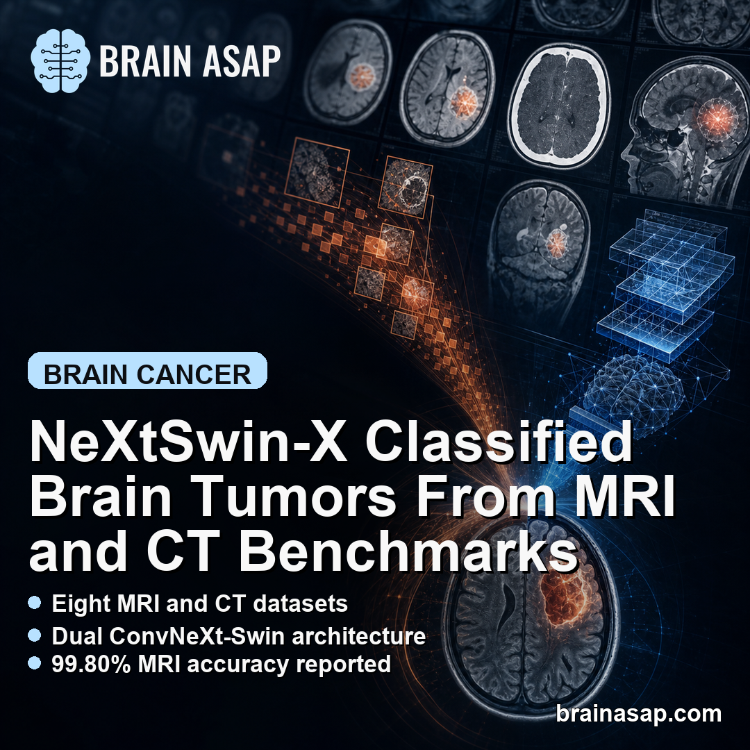
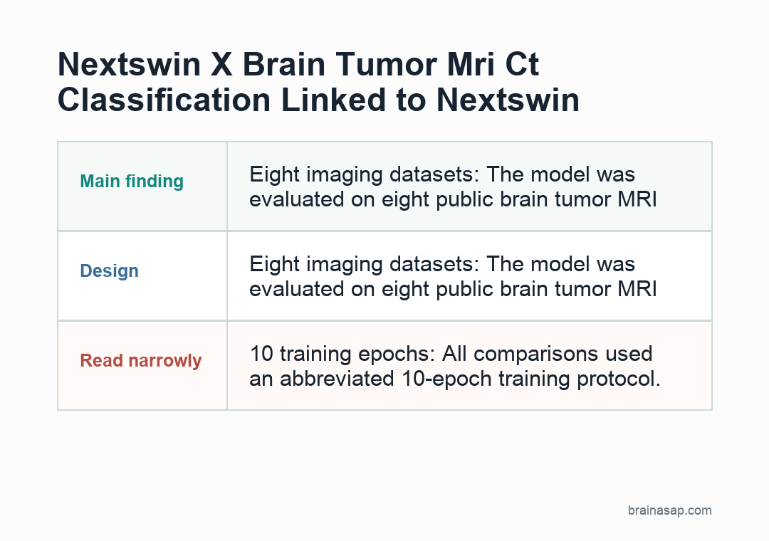
- Eight imaging datasets: The model was evaluated on eight public brain tumor MRI and CT datasets.
- Dual-branch architecture: NeXtSwin-X paired ConvNeXt for local image patterns with a Swin Transformer for global context.
- 10 training epochs: All comparisons used an abbreviated 10-epoch training protocol.
- 99.80% MRI accuracy: On the Brain Tumor Multimodal MRI subset, NeXtSwin-X reached 99.80% accuracy and F1-score.
- 99.78% CT accuracy: On the Brain Tumor Multimodal CT subset, the model reached 99.78% accuracy and F1-score.
Source: Scientific Reports (2026) | Esfahani et al.
NeXtSwin-X is an artificial intelligence model for brain tumor image classification. It was designed for a common medical-AI problem: strong models can perform well, but fine-tuning large systems on modest medical datasets can be computationally expensive.
The paper’s claim is that combining complementary pretrained backbones can produce efficient adaptation across MRI and CT tumor datasets without long training schedules.
NeXtSwin-X Combined ConvNeXt and Swin Transformer Features
The model used a dual-branch design.
One branch used ConvNeXt, a modern convolutional neural network suited to local visual patterns. The other used a Swin Transformer, which captures broader context across image regions.
A cross-attention mechanism fused the two feature streams. The authors also used differential learning rates, allowing the newly added fusion and classification layers to learn more aggressively while pretrained backbones adapted more conservatively.
That training setup is meant to reduce wasted computation. Instead of forcing every layer to relearn from a small tumor dataset, the model reuses general image features and concentrates updating where the brain-tumor task needs the most adaptation.
- Local patterns: ConvNeXt can capture edges, textures, and lesion-level features in brain images.
- Global context: Swin Transformer can model wider spatial relationships that may help distinguish tumor classes.
- Transfer learning: Pretrained backbones reduce the need to train a large model from scratch.
That architecture is a plausible fit for neuroimaging classification because tumors can carry both local signal and broader anatomical context.
Eight MRI and CT Datasets Tested Generalization
The evaluation covered eight public datasets. MRI tasks included multi-class glioma, meningioma, pituitary tumor, and no-tumor classification, while CT tasks included binary tumor classification and a smaller multi-class abnormality dataset.
Dataset sizes ranged widely. The Nickparvar MRI dataset had 7,023 images, BRISC 2025 had 6,000, and the CT Brain Abnormality dataset had only 259 images.
- Large MRI benchmarks: Nickparvar, BRISC, Sartaj, Orvile/Rahman, and Br35H tested common MRI tumor-classification tasks.
- Multimodal MRI and CT: Separate 5,000-image MRI and 4,618-image CT subsets tested binary tumor-versus-healthy classification.
- Small CT benchmark: The 259-image CT abnormality dataset tested a harder small-data multi-class setting.
Using multiple datasets is important because single-dataset medical-AI results can look stronger than they are. Different sources can vary in scanners, preprocessing, class balance, and label quality.
Performance Was Strong After 10 Training Epochs
All model comparisons used 10 training epochs. The paper reported that validation accuracy and F1-score often leveled off by about epochs 4-6, suggesting rapid specialization.
Selected results were high. NeXtSwin-X reached 98.95% accuracy on Nickparvar, 98.90% on BRISC 2025, 99.33% on Br35H, 99.80% on the multimodal MRI subset, and 99.78% on the multimodal CT subset.
- Harder Sartaj result: Accuracy was lower at 80.96%, but still higher than the ConvNeXt-only result of 76.14%.
- Small CT result: The CT Brain Abnormality dataset reached 100.00% accuracy, which should be interpreted cautiously because the dataset had 259 images.
- Baseline comparison: NeXtSwin-X generally matched or outperformed single-backbone and ResNet-style baselines.
The weaker Sartaj result is useful because it prevents a simplistic “near-perfect AI” reading. Performance depended on dataset difficulty and curation.
AI Accuracy Does Not Equal Clinical Readiness
Brain tumor classification models can assist research and triage, but public benchmark performance is not the same as hospital deployment. Real clinical images include artifacts, atypical cases, prior surgery, treatment effects, and scanner variability.
The paper evaluated classification, not radiologist workflow integration, prospective safety, calibration, or patient outcomes.
A model can classify benchmark images well and still need substantial validation before clinical use.
- External validation: Hospital-held-out datasets are needed to test performance outside public benchmarks.
- Clinical labels: Tumor diagnosis may depend on imaging sequence, pathology, molecular markers, and longitudinal context.
- Workflow risk: False reassurance or false alarm can matter if AI output is treated as more certain than the clinical evidence supports.
The most defensible interpretation is technical. NeXtSwin-X appears to be an efficient hybrid architecture for brain tumor image classification, not a stand-alone diagnostic tool.
The Main Advance Is Efficient Transfer Learning
The model’s practical appeal is computational efficiency. By using pretrained backbones and differential learning rates, the system adapted quickly across datasets rather than requiring prolonged full-model training.
That could matter for medical imaging groups with limited compute or modest labeled datasets. Efficient fine-tuning can make model development more accessible, provided validation standards remain strict.
- Engineering result: Cross-attention fusion helped combine convolutional and transformer features.
- Benchmark result: Accuracy and F1 were high on most MRI and CT datasets.
- Clinical next step: Prospective, institution-independent testing is needed before any patient-facing use.
For brain-health readers, the important point is that medical AI is moving toward hybrid systems that combine local image detail with broader anatomical context. The scientific question now shifts from benchmark accuracy to reliable clinical generalization.
Citation: DOI: 10.1038/s41598-026-50158-1. Esfahani et al. NeXtSwin-X: dual-branch cross-attention fusion of ConvNeXt and Swin Transformer for accurate brain tumor classification from MRI and CT. Scientific Reports. 2026.
Study Design: Technical validation study across public MRI and CT brain tumor image datasets.
Sample Size: Eight public datasets totaling tens of thousands of MRI and CT images across binary and multi-class tasks.
Key Statistic: NeXtSwin-X achieved 99.80% accuracy on the multimodal MRI subset and 99.78% on the multimodal CT subset after 10 epochs.
Caveat: Public benchmark performance does not establish prospective clinical readiness or diagnostic safety.