TL;DR: A 2026 meta-analysis in Psychological Medicine found that reduced resting-state connectivity between the amygdala and anterior cingulate cortex was the most robust brain-network difference in anxiety disorders.
Key Findings
- 15 datasets: The meta-analysis pooled 14 studies with 15 independent datasets of amygdala-based resting-state functional MRI.
- 783 total participants: Included samples covered 378 people with anxiety disorders and 405 healthy controls.
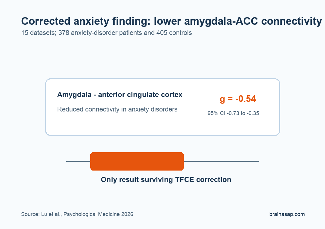
- Corrected ACC result: After threshold-free cluster enhancement correction, only reduced amygdala-anterior cingulate cortex connectivity remained significant.
- Effect size g = -0.54: The corrected amygdala-ACC reduction had a 95% confidence interval from -0.73 to -0.35.
- Left-amygdala pattern: Subgroup analyses suggested that the corrected result was driven mainly by adults and the left amygdala.
Source: Psychological Medicine (2026) | Lu et al.
Resting-state functional connectivity measures which brain regions show coordinated activity while a person is not doing a specific task. Anxiety studies have often focused on the amygdala because it helps process threat, fear, and emotional salience.
Individual imaging studies have not always agreed. Some reported stronger amygdala-prefrontal connections in anxiety, others reported weaker connections, and many were small.
This meta-analysis tested which amygdala network abnormality survives across studies and correction methods.
Meta-Analysis Pooled Amygdala Resting-State MRI Across Anxiety Disorders
Researchers searched Embase, PubMed, and Web of Science through December 26, 2025. Eligible studies compared people with anxiety disorders against healthy controls using whole-brain resting-state functional magnetic resonance imaging (fMRI) with the amygdala as the seed region.
The analysis focused on social anxiety disorder, generalized anxiety disorder, and separation anxiety disorder. It excluded studies of post-traumatic stress disorder, obsessive-compulsive disorder, panic disorder, specific phobia, and subthreshold anxiety.
- Initial search: 2,595 original or review records were identified before screening.
- Full-text review: 101 studies were evaluated in detail.
- Final pool: 14 studies contributed 15 independent datasets.
- Sample size: The final analysis included 378 anxiety-disorder patients and 405 healthy controls.
The statistical method was Seed-based d Mapping with Permutation of Subject Images, or SDM-PSI. That approach uses voxel-wise tests and correction procedures meant to reduce false-positive neuroimaging findings.
The stricter correction step was threshold-free cluster enhancement, a method designed to identify spatially coherent clusters without relying only on one arbitrary cluster-forming threshold.
Amygdala-ACC Connectivity Was the Corrected Anxiety Signal
The clearest result was lower connectivity between the amygdala and the anterior cingulate cortex (ACC). The ACC is involved in emotion regulation, conflict monitoring, attention, and integration between emotional and cognitive control systems.
Before the strictest correction, anxiety-disorder datasets showed several amygdala-connectivity differences. After threshold-free cluster enhancement correction, the amygdala-ACC reduction was the only result that remained significant.
- Corrected decrease: Amygdala-ACC connectivity was lower in anxiety disorders than in controls, with g = -0.54.
- Confidence interval: The 95% confidence interval ran from -0.73 to -0.35.
- Corrected p value: The corrected result was reported at p = 0.001.
- Cluster location: The corrected cluster was centered in the left ACC and extended into the right ACC.
Lower amygdala-ACC connectivity fits a common model of anxiety in which regulatory circuits do not coordinate normally with threat-processing circuits.
The pooled finding does not prove that weak ACC control causes anxiety. It identifies the most reproducible imaging abnormality in the available amygdala-seeded resting-state literature.
Temporal and Visual Cortex Results Were Less Robust
Uncorrected analyses also found higher amygdala connectivity with the left superior temporal gyrus, middle temporal gyrus, and cuneus. Those regions are involved in social perception, auditory/language processing, visual processing, and broader sensory integration.
The temporal and cuneus clusters appeared only before the stricter correction step. They should be read as less robust than the amygdala-ACC decrease.
- Superior temporal gyrus: Increased connectivity had g = 0.46, with a 95% confidence interval from 0.27 to 0.65.
- Middle temporal gyrus: Increased connectivity had g = 0.38, with a 95% confidence interval from 0.19 to 0.57.
- Cuneus: Increased connectivity had g = 0.35, with a 95% confidence interval from 0.17 to 0.53.
This distinction is important for biomarker language. A pooled uncorrected map can show a cluster that is still too fragile to carry the central circuit interpretation.
Adult and Left-Amygdala Analyses Supported the ACC Connectivity Decrease
Subgroup analyses suggested that the amygdala-ACC result was driven mainly by adult patients and by the left amygdala. The adult-only analysis included 11 datasets and produced patterns largely consistent with the full analysis.
Left-right differences are plausible because amygdala function is not always symmetrical. Still, subgroup analyses are usually less stable than main analyses, so the left-amygdala point should be read as a directional clue rather than a finished lateralization rule.
The sensitivity tests strengthened confidence in the corrected amygdala-ACC decrease. Jackknife analysis, which repeats the meta-analysis while leaving out one dataset at a time, found that amygdala-left ACC connectivity survived correction in 12 of 15 study combinations.
Amygdala-ACC Connectivity Is a Research Target, Not a Diagnostic Test
The meta-analysis supports a focused claim: across seed-based resting-state fMRI studies, anxiety disorders most consistently showed lower amygdala-ACC connectivity. That is different from saying a scan can diagnose anxiety in an individual patient.
The included studies varied in diagnosis, age, scanner methods, medication status, symptom scales, and analytic thresholds. Meta-regression did not find significant correlations with age, sex distribution, or anxiety severity, but study-level analyses have limited power.
For treatment research, the amygdala-ACC circuit remains a plausible target for studies of psychotherapy, medication, neuromodulation, and biomarkers. For clinical care today, the corrected connectivity decrease is better used to understand anxiety circuitry than to guide individual treatment decisions.
Citation: DOI: 10.1017/S0033291726104310. Lu et al. Altered amygdala resting-state functional connectivity in anxiety disorders: a coordinate-based meta-analysis. Psychological Medicine. 2026.
Study Design: Coordinate-based meta-analysis of amygdala-seeded resting-state fMRI studies in anxiety disorders.
Sample Size: 15 datasets with 378 anxiety-disorder patients and 405 healthy controls.
Key Statistic: Reduced amygdala-ACC connectivity remained significant after correction, with g = -0.54 and 95% CI -0.73 to -0.35.
Caveat: The finding is a group-level neuroimaging marker, not an individual diagnostic test for anxiety disorders.