TL;DR: A 2026 cellular neuroscience study in iScience found that GPR3, a constitutively active G-protein-coupled receptor, behaved like an immediate-early gene and helped connect CREB signaling to presynaptic maturation during neuronal differentiation.
Key Findings
- Biphasic GPR3 induction: Gpr3 rose quickly after nerve growth factor stimulation, dipped, then increased again during later neuronal differentiation.
- NET-CAGE promoter readout: Native elongating transcript-cap analysis of gene expression detected activity at a core promoter about 200 base pairs upstream of the Gpr3 transcription start site.
- Five CRE sites tested: Five cAMP response elements in the 1-kb regulatory region cooperated to support stimulus-responsive Gpr3 transcription.
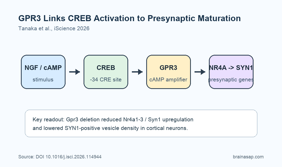
- Proximal CREB binding: Phosphorylated CREB was enriched at the proximal -34 cAMP response element, linking cAMP signaling to early Gpr3 transcription.
- Synapsin pathway reduced by deletion: In primary cortical neurons, Gpr3 deletion weakened developmental Nr4a1-3 and Syn1 upregulation and reduced SYN1-positive vesicle density.
Source: iScience (2026) | Tanaka et al.
GPR3 is a Gs-coupled receptor that can raise intracellular cyclic AMP even without a known ligand. In this setting, the receptor acted as a standing amplifier inside cells, not only as a detector of an outside signal.
The research team tested whether this receptor also behaves like a stimulus-responsive neuronal gene. Their experiments used PC12 cells, a standard rat cell model for nerve growth factor-driven neuronal differentiation, and primary mouse cortical neurons.
GPR3 Rose Early and Again Later During Neuronal Differentiation
Researchers first tracked Gpr3 mRNA after nerve growth factor (NGF) pushed PC12 cells toward a neuronal state. The response was not a slow single increase.
Instead, Gpr3 showed a biphasic expression pattern: an early rise, a decline, and then a later sustained increase. That timing made it resemble immediate-early genes, the fast-response genes neurons use to translate activity into longer-term changes.
The cell model showed the expected differentiation markers. NGF and forskolin increased beta-III tubulin and synapsin I by 48 hours, while potassium chloride produced weaker differentiation signals.
The time course matters because GPR3 was not just present in differentiating cells. It moved during the transition, which supports a regulatory role in the differentiation program.
- Early phase: Gpr3 increased rapidly after NGF and cAMP stimulation, placing it near the fast-response part of the gene-expression cascade.
- Late phase: Gpr3 increased again during the period when neuronal markers and synaptic proteins were rising.
- Model boundary: PC12 cells are useful for neuronal differentiation logic, but they are not a full brain circuit.
CREB Binding Linked cAMP Signaling to the Gpr3 Promoter
The team used NET-CAGE, a method that captures nascent capped RNA, to locate active transcription during NGF-induced differentiation. The signal pointed to a core promoter around 200 base pairs upstream of the Gpr3 transcription start site.
At 30 minutes after NGF stimulation, Gpr3 showed roughly 10-fold induction in the NET-CAGE readout and ranked 27th among upregulated genes. The researchers noted that no other G-protein-coupled receptor showed a similar magnitude of induction.
The promoter analysis then narrowed the mechanism. The 1-kb upstream regulatory region contained five cAMP response elements, and reporter experiments showed that those elements cooperated to mediate stimulus-responsive transcription.
Phosphorylated CREB, the activated form of cAMP response element-binding protein, was enriched at the proximal -34 CRE. This connects Gpr3 induction to a familiar neuronal pathway: cAMP signaling activates CREB, and CREB helps turn on genes involved in neuronal adaptation.
GPR3 Reinforced the NR4A-Synapsin Presynaptic Program
After mapping the promoter response, researchers tested whether GPR3 affected downstream neuronal genes. The main pathway involved Nr4a1-3, a group of activity-responsive nuclear receptor genes, and Syn1, the gene encoding synapsin I.
In PC12 cells, early Gpr3 induction enhanced delayed Nr4a1-3 expression. The experiments also supported an Nr4a1-dependent route from GPR3 signaling to Syn1 transcription.
That pathway is biologically coherent. GPR3 can raise cAMP tone; cAMP can feed CREB signaling; CREB-responsive NR4A factors can then help regulate genes involved in neuronal differentiation and synaptic development.
The tested sequence had four steps:
- Stimulus arrives: NGF or cAMP-linked stimulation activates early transcriptional machinery.
- Gpr3 turns on: CREB-linked promoter elements support fast Gpr3 induction.
- cAMP tone is reinforced: GPR3 acts as an activity-dependent amplifier inside the differentiating cell.
- Presynaptic markers rise: NR4A signaling supports Syn1 expression and presynaptic maturation.
Primary Cortical Neurons Showed Lower Synapsin-Positive Vesicle Density
The strongest neural relevance came from primary cortical neurons. When Gpr3 was deleted, the developmental upregulation of Nr4a1-3 and Syn1 was diminished.
The deletion also reduced SYN1-positive vesicle density. Synapsin I is associated with presynaptic vesicles, so this finding ties the transcriptional pathway to a structural marker of presynaptic development.
GPR3 is only one part of synapse development. Neuronal differentiation uses many overlapping signals, including growth factors, membrane depolarization, cAMP pathways, calcium signaling, and activity-dependent transcription factors.
The narrower interpretation is stronger: GPR3 helps sustain part of the activity-linked transcriptional program that supports presynaptic maturation.
- Cell-line evidence: PC12 experiments defined the timing and promoter mechanism.
- Primary-neuron evidence: Cortical neuron deletion showed reduced NR4A/Syn1 upregulation and fewer SYN1-positive vesicles.
- Translation limit: The work did not test cognition, behavior, or disease treatment in animals or humans.
GPR3 Is a Mechanism Target, Not a Treatment Claim
Immediate-early gene systems are important because they help neurons convert experience into cellular change. This study adds GPR3 to that type of logic, but as a receptor-like amplifier rather than a classic transcription factor.
The work also gives researchers a specific pathway to test in disease models. GPR3 has been discussed in neuronal biology before, and immediate-early gene dysregulation has been linked to neurodevelopmental and cognitive disorders, but this experiment stayed at the cellular mechanism level.
The evidence supports GPR3 as an activity-dependent cAMP amplifier during neuronal differentiation. It does not establish that changing GPR3 would improve memory, autism traits, neurodegeneration, or any clinical condition.
The useful next experiments would test whether this CREB-GPR3-NR4A-Syn1 pathway changes in intact developing brain circuits, whether it affects synaptic function, and whether disease-linked perturbations alter the same sequence.
Citation: DOI: 10.1016/j.isci.2026.114944. Tanaka et al. GPR3 is an immediate-early gene-like GPCR regulating CREB-dependent neuronal differentiation. iScience. 2026;29:114944.
Study Design: Cellular and molecular neuroscience study using PC12 neuronal differentiation experiments, NET-CAGE promoter mapping, reporter assays, gene perturbation, and primary cortical neuron validation.
Sample/Model: Rat PC12 cells and primary mouse cortical neurons.
Key Statistic: Gpr3 showed approximately 10-fold induction at 30 minutes after NGF stimulation in the NET-CAGE readout and ranked 27th among upregulated genes.
Caveat: The experiments identify a neuronal differentiation mechanism in cell models, not a behavioral or clinical effect in living animals or patients.