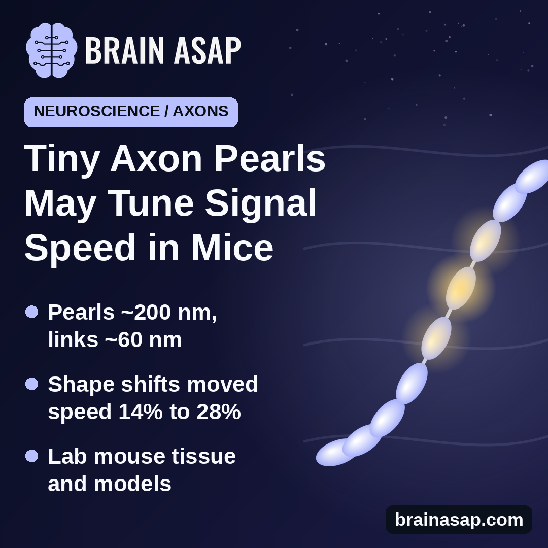
TL;DR: Thin nerve fibers in the mouse brain are not smooth tubes but look like strings of tiny pearls, a shape set by the physics of their outer membrane. Changing that shape with drugs or heavy activity sped up or slowed down nerve signals by roughly 14% to 28% in mouse brain slices.
Key Findings
- Pearls on a string: Swellings about 200 nanometers wide joined by links about 60 nanometers wide.
- Hidden by standard methods: Seen with ultra-fast freezing, but not in chemically fixed tissue.
- Membrane physics: Salt balance, cholesterol and a motor protein all changed the pearls.
- Shape changed speed: Removing cholesterol slowed signals about 28%; blocking the motor protein sped them up 19%.
- Activity reshaped axons: Bursts of stimulation enlarged the pearls and cut membrane cholesterol by about 45%.
Source: Nature Neuroscience (2025) | Griswold et al.
Textbook drawings show axons, the long wires that carry nerve signals, as smooth cables. A team led from Johns Hopkins University and the University of California, San Diego found that the thinnest ones look more like a string of pearls.
The study was published online in December 2024. It suggests the shape of these fibers is set largely by simple membrane physics, and that the shape changes how fast signals travel.
Why No One Saw the Pearls Before
Brain tissue is usually prepared for electron microscopy with chemical fixatives, which can distort fine structures. In that tissue, thin axons look like even tubes.
The researchers instead froze mouse brain tissue in milliseconds under high pressure, which preserves its natural shape. They studied three kinds of samples:
- Fresh brain tissue: From adult mice
- Brain slices kept in culture: For weeks
- Neurons grown in dishes: From embryonic mice
In all three, thin axons without an insulating myelin coat showed the same pattern: small swellings about 200 nanometers wide, linked by stretches as thin as 60 nanometers. A human hair is roughly 80,000 nanometers across.
These swellings are not synapses, where neurons connect, and they are far smaller than the large beads seen on dying axons.
Membrane Physics Shapes the Pearls
Thin tubes made of fatty membrane tend to break up into beads under tension, a known effect in physics. Computer models of axon membranes reproduced the pearls, and experiments backed them up:
- Salt balance: Changing the saltiness of the surrounding fluid changed pearl size
- Cholesterol: Stripping it from the membrane made pearls rounder
- Motor protein: Blocking myosin II, which tightens the cell’s inner scaffold, widened the thin links
Pearl Shape Changed Signal Speed
Models predicted that wider links and well-proportioned pearls would let signals travel faster. Recordings in mouse brain slices agreed.
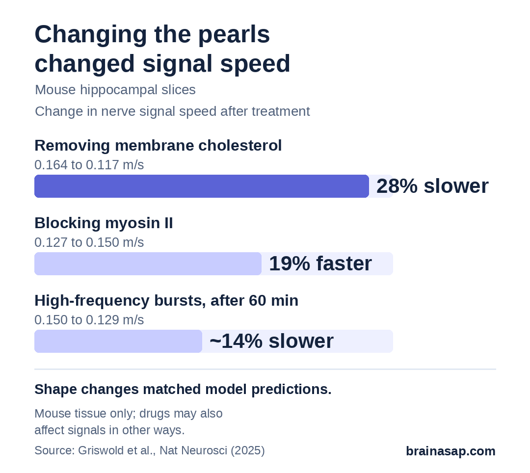
Because cholesterol removal could affect signals in other ways, the team also used the myosin blocker. It changed shape without stripping cholesterol, and it sped signals up as the model predicted.
Busy Axons Change Shape
The researchers then gave neurons three bursts of rapid stimulation. Within 5 minutes, the pearls grew about 8% longer and 17% wider, while membrane cholesterol dropped by about 45%.
Over the next hour, signals in brain slices slowed from 0.150 to 0.129 meters per second. In slices already stripped of cholesterol, the speed did not change in the same way, pointing to cholesterol as part of the mechanism.
Basic Biology, Not a Health Claim
- Mouse tissue only: Human axons were not examined
- Thin, uninsulated axons: The thick, myelin-wrapped fibers were not the focus
- Lab conditions: Sizes varied with how tissue was prepared
- Models plus slices: Speed effects come from simulations and brain-slice recordings, not living animals
- Drugs have side effects: Cholesterol stripping and myosin blockers may affect signals in other ways
Nothing here says that dietary cholesterol or any supplement changes how fast you think.
A New Way Axons Might Fine-Tune Timing
The finding adds a new layer to how the brain could adjust the timing of its signals: not only through insulation or synapses, but through the shape of the wire itself. The next question is whether nanopearls behave the same way in living brains and in human tissue, and whether they change in disease.
Citation: DOI: 10.1038/s41593-024-01813-1. Griswold JM, Bonilla-Quintana M, Pepper R, et al. Membrane mechanics dictate axonal pearls-on-a-string morphology and function. Nat Neurosci. 2025;28:49-61.
Study Design: High-pressure-freezing electron microscopy, membrane modeling and electrophysiology in mouse brain tissue, slice cultures and cultured neurons.
Sample Size: Multiple preparations; for example, 300 axons from 3 independent cultures per condition and 12 slices per drug condition in recordings.
Key Statistic: Conduction velocity fell from 0.164 to 0.117 m/s after cholesterol removal (~28%) and rose from 0.127 to 0.150 m/s with blebbistatin (19%).
Caveat: Mouse tissue and models only; unmyelinated axons; drug effects not purely structural.