TL;DR: In 86 people from one hospital in China, those with Parkinson’s and mild thinking problems showed changes in how water moves through parts of the hippocampus, a memory center. The hippocampus was not measurably smaller, suggesting a diffusion scan may pick up changes that size alone misses.
Key Findings
- No size difference: None of 38 hippocampal volume measures differed between groups.
- Water movement differed: 42 diffusion measures did, after correcting for 182 tests.
- Linked to thinking problems: Most differences separated Parkinson’s with thinking problems from both other groups.
- One early sign: One measure was already lower in Parkinson’s with normal thinking.
- Not a test yet: Links to thinking scores vanished once group was accounted for.
Source: Frontiers in Aging Neuroscience (2026) | Chen et al.
Many people with Parkinson’s develop problems with thinking and memory, and those problems can be an early step toward dementia. Doctors would like a scan that flags trouble early, but standard MRI often shows nothing obvious.
Neurologists at the First People’s Hospital of Lianyungang in China looked closely at the hippocampus, a seahorse-shaped memory center, using two kinds of MRI.
Three Groups and Two Kinds of Scan
The study looked back at scans from 86 people:
- Parkinson’s with mild thinking problems (34): Lower scores on memory and thinking tests, but not dementia
- Parkinson’s with normal thinking (34): Similar disease length, stage and medication
- Healthy adults (18): Similar age and sex
Mild thinking problems were defined with the Movement Disorder Society’s simpler (Level I) criteria. The two Parkinson’s groups had median scores of 20 and 28 on the 30-point MoCA thinking test.
The researchers measured 19 small subfields on each side of the hippocampus in two ways:
- Volume: How big each subfield was
- Diffusion: How freely water moved through the tissue, which tends to rise when tissue structure loosens or breaks down
The Hippocampus Was Not Smaller
After accounting for age, sex, education and head size, none of the 38 volume measures differed significantly between groups. Total hippocampus volume did not either.
The authors note the study had limited power to find small or moderate size differences.
Water Moved More Freely in Parkinson’s With Thinking Problems
Diffusion told a different story. Of 144 diffusion measures, 42 differed between groups after correcting for all 182 tests. Water moved more freely in many subfields in people with thinking problems, with the strongest effect in a part of the hippocampus called the dentate gyrus.
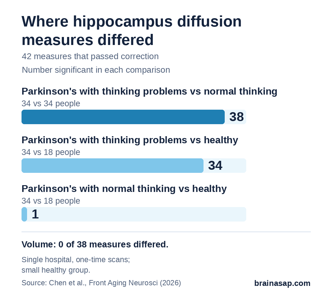
That single early difference, present even in people with normal thinking, hints that some changes come with Parkinson’s itself rather than with thinking problems.
Linked to Group, Not Clearly to Test Scores
Across everyone with Parkinson’s, 39 of the 42 diffusion measures tracked MoCA scores. But once the researchers accounted for which group each person was in, none of those links held.
That means the scans separated the groups but did not clearly add information about a person’s exact thinking score.
A Snapshot With Technical Limits
- One hospital, looking back: Records from a single center
- Small healthy group: Only 18 people
- One point in time: Cannot show whether changes predict decline
- Coarse images: Diffusion scans at 2 mm, subfields drawn from standard 1 mm images without a dedicated high-resolution sequence
- Uncorrected distortion: Some image warping was not corrected
- Indirect measure: Water diffusion does not directly show nerve damage or myelin loss
- Education differed: Adjusted for, but groups were not matched
Follow-Up Scans Over Time Are the Next Step
The results suggest diffusion MRI may catch hippocampal changes in Parkinson’s before shrinkage shows up. The question for the next study is whether these changes, measured over several years in more people, predict who goes on to develop dementia.
Citation: DOI: 10.3389/fnagi.2026.1946629. Chen Y, Peng L, Hu Q, Zhao Z, Xu B. Hippocampal subfield diffusion abnormalities in Parkinson’s disease with mild cognitive impairment: a volumetric and diffusion MRI study. Front Aging Neurosci. 2026.
Study Design: Retrospective cross-sectional single-center MRI study.
Sample Size: 34 Parkinson’s with mild cognitive impairment, 34 Parkinson’s with normal cognition, 18 healthy controls.
Key Statistic: 0 of 38 volume measures and 42 of 144 diffusion measures differed after FDR correction across 182 outcomes.
Caveat: Small single-center snapshot; limited image resolution; MoCA links disappeared after adjusting for group.