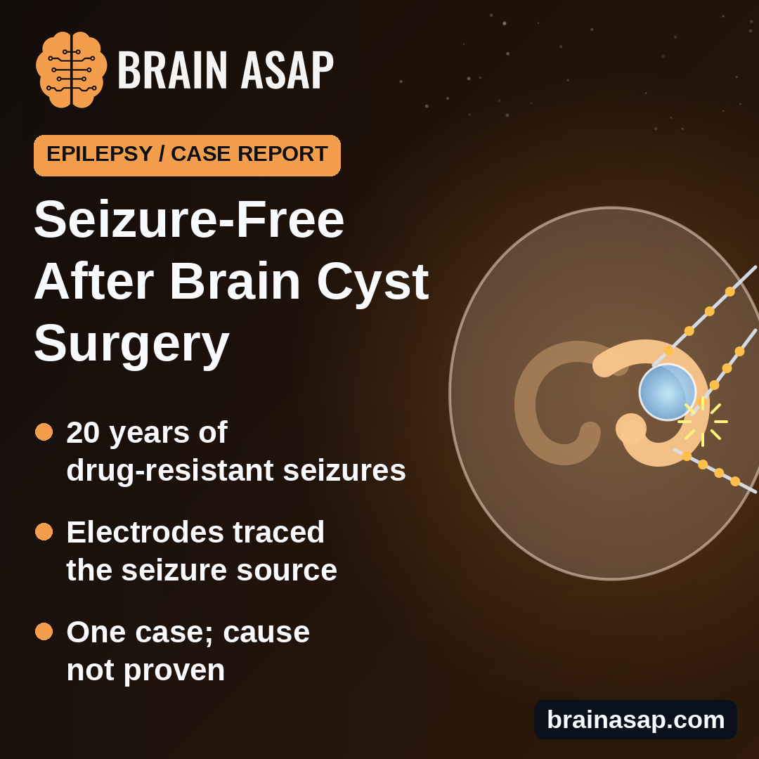
TL;DR: A 35-year-old woman with 20 years of seizures that medicines could not control became seizure-free after surgery removed a small fluid-filled cyst and the nearby part of her brain where seizures started. It is one case, so it cannot prove the cyst caused her epilepsy.
Key Findings
- Seizures since age 15: Up to 2 to 3 a week despite many drugs.
- 1.2 cm cyst: Sat right next to the memory-related hippocampus.
- Brain electrodes found the source: 12 of 15 recorded seizures began in the hippocampus area beside the cyst.
- Seizure-free at 12 months: After heat treatment, then surgery removing the cyst and hippocampus.
- Mood improved: Depression score fell from 32 to 9.
Source: Frontiers in Neuroscience (2026) | Fu, Ye et al.
Brain scans often turn up small fluid-filled cysts that doctors consider harmless. One type, called a choroidal fissure cyst, sits deep in the temporal lobe near the hippocampus, a structure that is also a common starting point for seizures. Whether these cysts can actually cause epilepsy has been debated for decades.
A team at Shandong First Medical University in China describes a patient where the case for a link is unusually detailed.
Twenty Years of Seizures Drugs Couldn’t Stop
The woman’s first seizure came at 15, after a high fever. Over time she developed nighttime episodes of sitting up, fumbling with her hands and calling out without awareness, plus daytime falls and full convulsions. At their worst they came 2 to 3 times a week.
She tried carbamazepine, oxcarbazepine, valproate, lamotrigine, clonazepam, lacosamide and zonisamide. None brought her seizures under control. Before surgery, her scores showed marked anxiety, moderate depression and mild memory and thinking problems.
A Small Cyst Beside the Hippocampus
MRI showed a 1.2 by 1.2 cm cyst on the right side, just outside the hippocampus and nudging it slightly. It looked like fluid, with no signs of a tumor or swelling. The hippocampus beside it was a little smaller than normal but lacked the scarring that often causes this kind of epilepsy. A PET scan showed lower activity in the right hippocampus and amygdala.
Electrodes Inside the Brain Traced the Seizures
To find exactly where seizures started, surgeons placed thin recording electrodes deep in the brain, a technique called SEEG. Of 15 seizures recorded, 12 began in the right hippocampus and the area around it, next to the cyst. Mild electrical stimulation of the right hippocampus recreated her usual warning sensation.
The team then used heat delivered through the electrodes to destroy the key spots. She had no seizures in the month that followed.
Surgery, Then a Seizure-Free Year
Because the cyst might keep irritating the area, surgeons then removed the front of the right temporal lobe, including the cyst, the front and middle of the hippocampus and nearby tissue. Tissue tests found no clear scarring or malformation.
Twelve months later she remained seizure-free on two medicines instead of five, and her EEG showed no significant abnormalities.
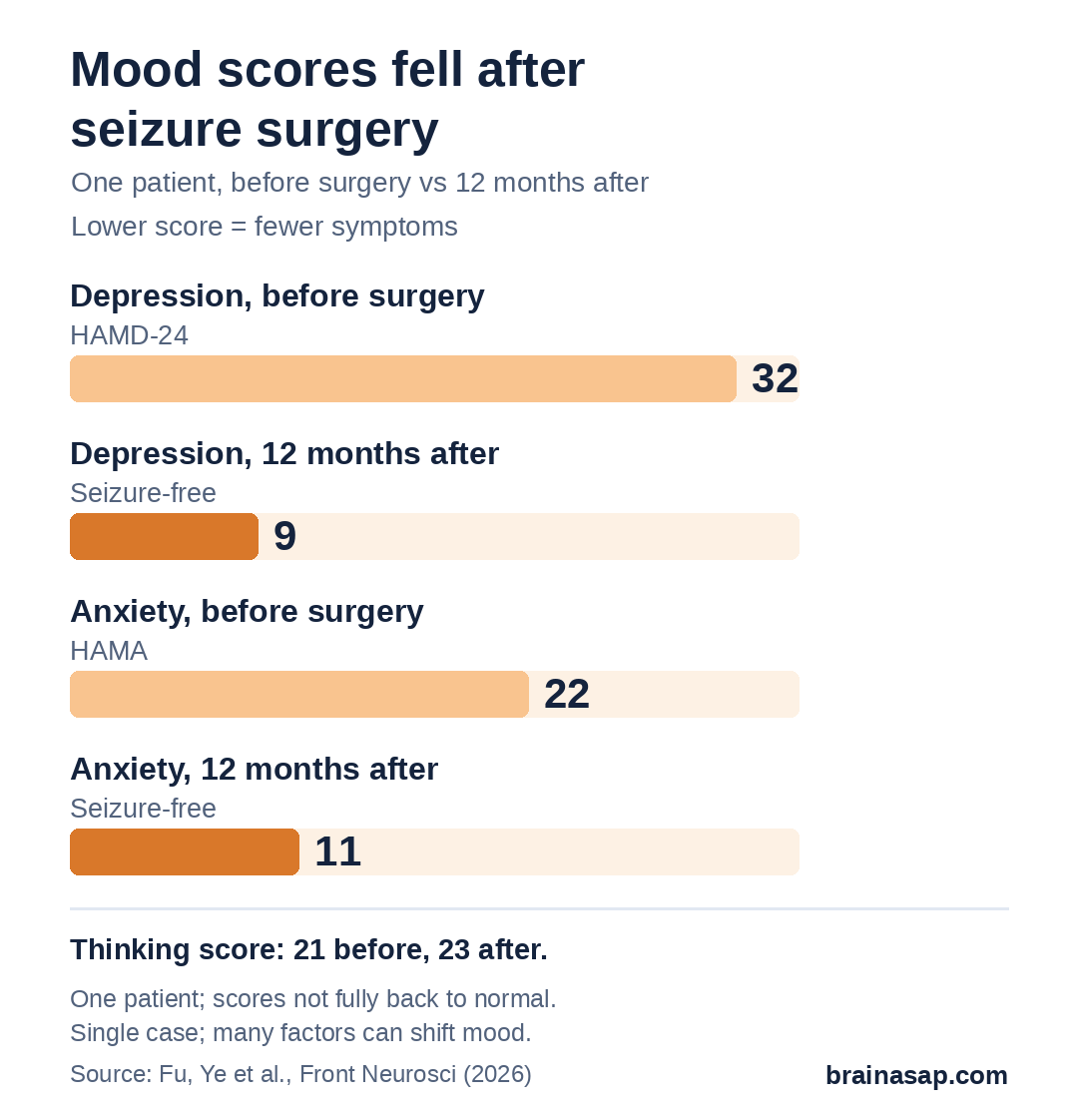
Her depression score dropped from 32 to 9 and her anxiety score from 22 to 11, though neither fully returned to normal. A thinking test score rose slightly, from 21 to 23, still in the mild impairment range.
Why One Case Can’t Prove the Cyst Was the Cause
- Single patient: No comparison group.
- More than the cyst was removed: Surgery also took out the hippocampus where seizures began.
- Seizures stopped before surgery: The earlier heat treatment alone was followed by a seizure-free month.
- Incomplete recordings: Electrodes did not capture every seizure type seen before.
- Short follow-up: One year so far.
Should Doctors Look Twice at “Harmless” Cysts?
For people with hard-to-treat epilepsy and a cyst near the hippocampus, this case suggests the cyst and its surroundings deserve a careful look rather than being dismissed. It does not mean the many people with such cysts and no seizures need treatment.
Whether removing the cyst alone, without the hippocampus, could stop seizures is the question that larger studies would need to answer.
Citation: DOI: 10.3389/fnins.2026.1905779. Fu G, Ye F, Lu S, Sun X, Zhou P. Case Report: Seizure freedom after resection of a temporal choroidal fissure cyst in drug-resistant epilepsy. Front Neurosci. 2026;20:1905779.
Study Design: Single case report with video-EEG, MRI, PET, SEEG, radiofrequency thermocoagulation and anterior temporal lobectomy.
Sample Size: 1 patient.
Key Statistic: Seizure-free at 12 months (Engel IA, ILAE Class 1).
Caveat: One case; surgery removed the hippocampus as well as the cyst; causality not established.