TL;DR: A 2026 preprint in medRxiv found that routine non-contrast brain computed tomography (CT) had a low acute-pathology yield in 291 emergency patients with new dizziness, while diabetes, ataxic gait, and headache marked higher odds of abnormal CT findings.
Key Findings
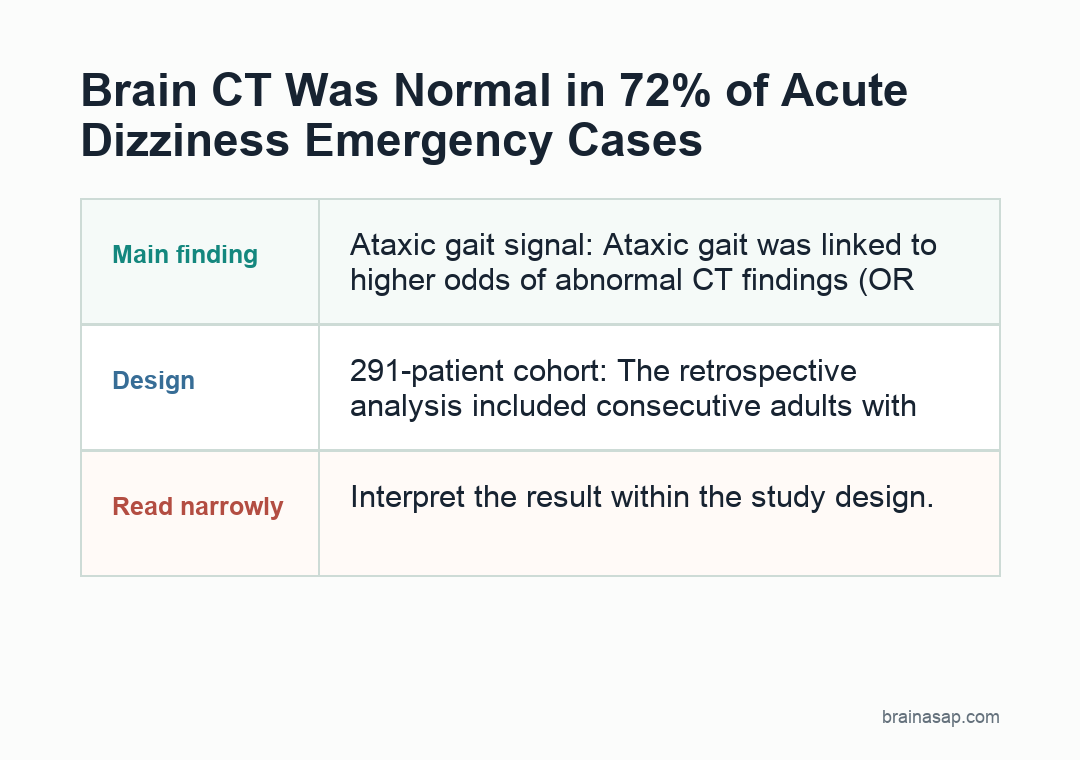
- 291-patient cohort: The retrospective analysis included consecutive adults with new-onset dizziness who underwent non-contrast brain CT at a tertiary emergency department.
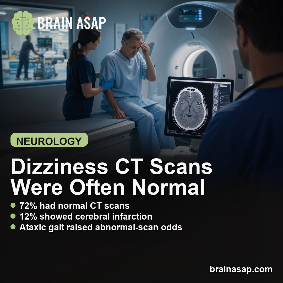
- 72.2% normal scans: CT showed no acute pathology in 210 of 291 patients, while 81 patients had abnormal findings.
- 12.0% cerebral infarction: The most common abnormal CT category was cerebral infarction, followed by neoplasm in 10.3% of the total cohort.
- Ataxic gait signal: Ataxic gait was linked to higher odds of abnormal CT findings (OR 2.41, 95% CI 1.18-4.93; q = 0.028).
- Nausea/vomiting contrast: Nausea (OR 0.45) and vomiting (OR 0.48) were associated with lower odds of abnormal CT findings after correction.
Source: medRxiv (2026) | Abbasi et al.
Acute dizziness is common in emergency care, but it can mean very different things. A benign vestibular syndrome, stroke, hemorrhage, tumor, medication effect, dehydration, or metabolic problem can all sit behind the same complaint.
The study focused on one imaging decision: when patients with new dizziness receive non-contrast brain CT, which bedside features line up with abnormal findings?
Non-Contrast Brain CT Was Normal in 72.2% of Acute Dizziness Cases
The analysis included 291 consecutive adult patients who presented with new-onset dizziness at Namazi Hospital, a tertiary referral center, between 2019 and 2021. Every patient in the cohort underwent non-contrast brain CT.
Average age was 59.4 years, and 146 patients were male. The most common medical history variables were hypertension, diabetes mellitus, and coronary artery disease.
- Common symptoms: Nausea appeared in 34.4%, vomiting in 31.6%, headache in 29.6%, and ataxic gait in 12.4%.
- Normal CT: 210 patients, or 72.2%, had no acute CT abnormality.
- Abnormal CT: 81 patients, or 27.8%, had an abnormal scan.
- Disposition: 56.4% were discharged, 38.5% were admitted, 3.8% were transferred, and 1.4% died.
Among abnormal scans, the two largest categories were cerebral infarction in 12.0% of the full cohort and neoplasm in 10.3%. Intracranial hemorrhage accounted for 3.1%.
Diabetes, Ataxic Gait, and Headache Marked Higher CT-Abnormality Odds
The regression analysis identified several variables tied to abnormal CT findings. The strongest outcome-linked variable was final disposition: patients admitted or dying had 8.01 times the odds of an abnormal scan compared with discharged patients.
That disposition result is not a bedside screening rule, because admission decisions can reflect the scan result itself. The clinically relevant predictors are the features available during evaluation.
- Diabetes mellitus: Abnormal CT odds were higher when diabetes was present (OR 3.10, 95% CI 1.52-6.33; q = 0.004).
- Ataxic gait: Gait ataxia was associated with higher abnormal-CT odds (OR 2.41, 95% CI 1.18-4.93; q = 0.028).
- Headache: Headache was also associated with higher abnormal-CT odds (OR 1.83, 95% CI 1.03-3.25; q = 0.049).
- Valvular heart disease: This history variable had a high estimate (OR 4.75), but the subgroup was small.
Ataxic gait is the clearest neurological clue in the list. Dizziness plus gait ataxia is more concerning for central nervous system involvement than isolated nausea or vomiting.
Nausea and Vomiting Tracked Lower Abnormal-CT Odds
Nausea and vomiting went in the opposite direction. Nausea was associated with lower odds of abnormal CT findings (OR 0.45, 95% CI 0.24-0.84), and vomiting showed a similar pattern (OR 0.48, 95% CI 0.26-0.88).
The evidence supports a narrow interpretation. These symptoms cannot rule out stroke or other central causes.
In this dataset, they were more common in patients whose CT showed no acute pathology, which fits the clinical idea that many vestibular presentations are peripheral.
- Younger than 60: Nausea and vomiting were stronger predictors of normal CT findings in the subgroup analysis.
- Women: Nausea was associated with normal CT findings in women, while no individual symptom reached significance in men.
- Older than 60: Individual symptom predictors were weaker, and final disposition carried the significant association.
Those subgroup results should be treated as exploratory. They are useful for hypothesis generation, not enough to create a practice-changing decision rule.
Single-Center CT Data Cannot Replace Stroke-Focused Bedside Evaluation
The main limitation is that non-contrast CT is weak for early ischemic stroke, especially when symptoms are recent. A normal CT cannot exclude every central cause of dizziness.
The researchers also did not have systematic MRI or follow-up imaging for patients with normal CT findings. That means some early ischemic strokes or delayed diagnoses could have been missed.
- Retrospective design: Results depended on chart documentation and existing clinical practice.
- Single center: A tertiary referral hospital may not match community emergency departments.
- No onset timing: The interval from dizziness onset to CT was not documented.
- No long-term follow-up: Delayed stroke, mortality, or functional outcomes could not be measured.
For acute dizziness, CT yield was low overall, and risk stratification should pay close attention to focal neurological signs such as ataxic gait, vascular history such as diabetes, and associated headache.
Prospective validation with standardized bedside exams, MRI follow-up, and timing data is needed before turning these predictors into an imaging rule.
Citation: DOI: 10.64898/2026.06.25.26356549. Abbasi et al. Correlation Between Clinical Presentation and Brain CT Findings in Acute Dizziness: A Retrospective Cross-Sectional Analysis at a Tertiary Referral Center. medRxiv. 2026.
Study Design: Retrospective cross-sectional preprint using emergency department records and non-contrast brain CT findings.
Sample Size: 291 adults with new-onset dizziness who underwent non-contrast brain CT.
Key Statistic: 72.2% of CT scans were normal; diabetes, ataxic gait, and headache were associated with higher abnormal-CT odds.
Caveat: Non-contrast CT can miss early ischemic stroke, and the single-center retrospective design needs prospective validation.