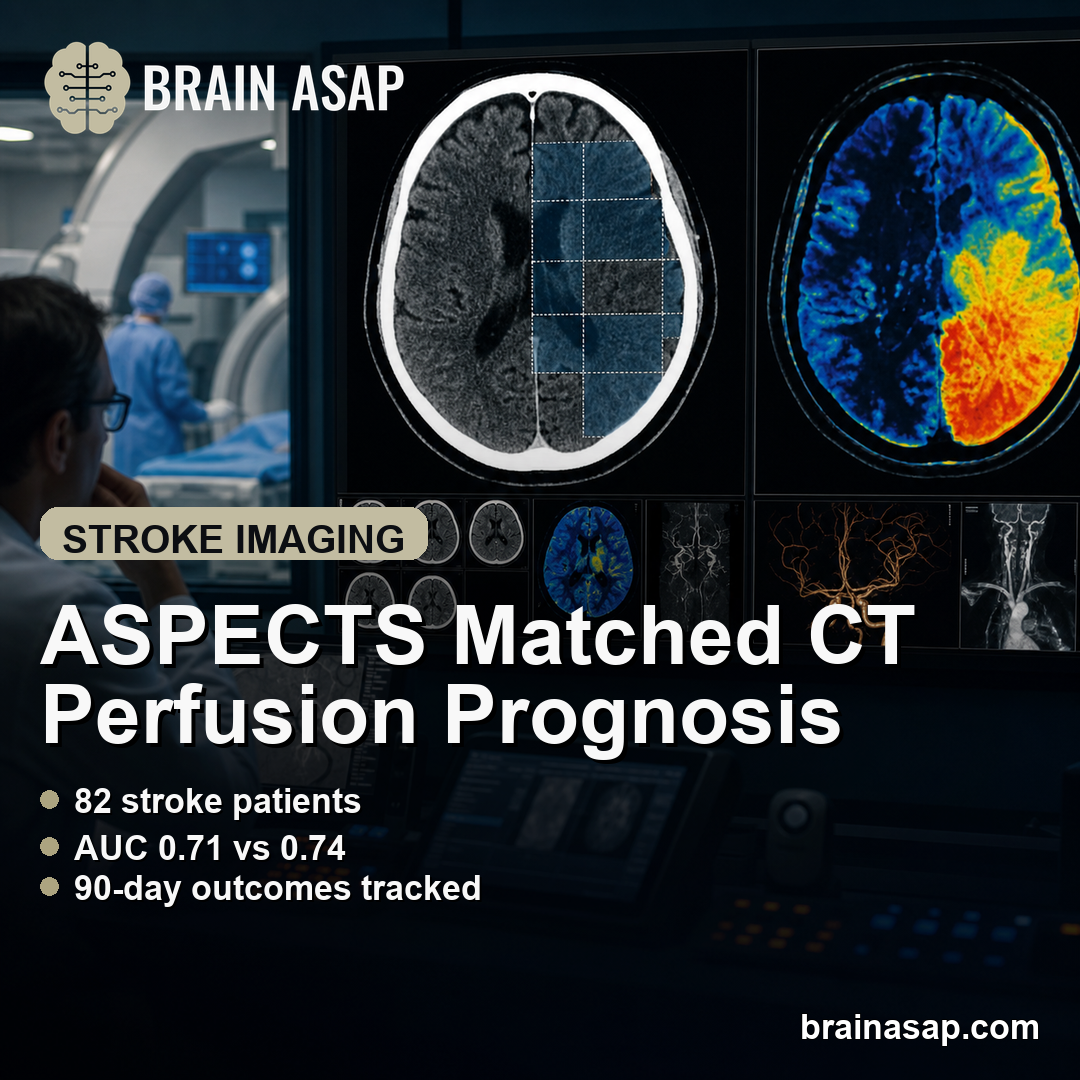
TL;DR: A 2026 study in the International Journal of General Medicine found that rapid ASPECTS scoring on non-contrast CT and CT perfusion core-infarct volume predicted 90-day outcomes similarly in 82 acute ischemic stroke patients treated with endovascular therapy.
Key Findings
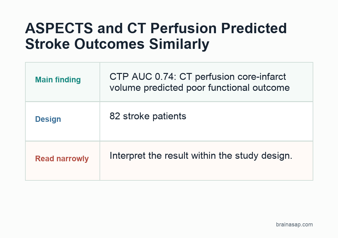
- 82 stroke patients: The retrospective single-center study included acute ischemic stroke patients treated with endovascular therapy within 24 hours.
- 45 poor outcomes: At 90 days, 45 patients had modified Rankin Scale scores above 2, while 37 had favorable outcomes.
- CTP AUC 0.74: CT perfusion core-infarct volume predicted poor functional outcome with an area under the ROC curve of 0.74.
- ASPECTS AUC 0.71: Directionally adjusted ASPECTS predicted poor outcome with an AUC of 0.71, not significantly different from CTP.
- Moderate inverse link: ASPECTS and CTP core volume were moderately negatively correlated, r = -0.61, p < .001.
Source: International Journal of General Medicine (2026) | Wang et al.
Acute ischemic stroke (AIS) treatment depends on speed and brain-tissue assessment. Clinicians need to know how much brain tissue already appears infarcted and whether imaging can help estimate the chance of recovery after thrombectomy.
Researchers compared 2 common imaging approaches: a quick non-contrast CT score called ASPECTS and a quantitative CT perfusion estimate of infarct core volume.
ASPECTS and CT Perfusion Both Predicted 90-Day Stroke Outcome
The study included 82 acute ischemic stroke patients treated at a single center between January 2023 and December 2024. All underwent endovascular therapy through an emergency stroke pathway.
Patients had anterior-circulation strokes, underwent both non-contrast CT and CT perfusion within 24 hours of symptom onset, and had 90-day functional outcome assessed with the modified Rankin Scale (mRS).
The cohort was clinically high risk. 72 patients had at least 1 comorbid condition, including hypertension in 57 patients, hyperglycemia in 28 patients, and atrial fibrillation in 24 patients.
Baseline neurological impairment was also meaningful. The median National Institutes of Health Stroke Scale (NIHSS) score was 12, while median ASPECTS was 9 and median CTP core volume was 20 mL.
The imaging comparison therefore occurred in a real thrombectomy workflow, not in a screening-only population. The analysis asked whether the fast score and the quantitative perfusion estimate offered similar prognostic information after patients had already reached endovascular treatment.
- Favorable outcome: mRS score of 2 or lower at 90 days.
- Poor outcome: mRS score above 2 at 90 days.
- Severe disability or death: mRS score of 4 or higher.
By that definition, 37 patients had favorable outcomes and 45 patients had poor outcomes. Patients with poor outcomes had larger CTP-derived infarct cores and lower ASPECTS scores, with both comparisons reported as p < .001.
The outcome endpoint was functional, not just radiologic. The analysis connected pre-treatment imaging to the patient’s level of disability or independence 3 months after therapy.
CTP Core Volume Had AUC 0.74 Versus ASPECTS AUC 0.71
The main comparison used receiver operating characteristic analysis, which asks how well a marker separates patients with poor outcomes from those with favorable outcomes. The metric was area under the curve, or AUC.
In the full cohort, CT perfusion (CTP) core-infarct volume had an AUC of 0.74. Directionally adjusted ASPECTS had an AUC of 0.71. The difference was not statistically significant.
CTP gave a quantitative infarct-core volume, but the simpler ASPECTS score still predicted outcome close to the CTP result.
- ASPECTS advantage: It is fast, widely available, and based on non-contrast CT.
- CTP advantage: It quantifies core infarct volume and can help characterize perfusion patterns.
- Clinical implication: The 2 measures may be complementary rather than interchangeable.
Lower ASPECTS Tracked Larger CTP Core Infarct Volume
The relationship between the two imaging measures went in the expected direction. Higher ASPECTS scores indicate less visible early ischemic injury, while larger CTP core volumes indicate more infarcted tissue.
The study reported a moderate negative correlation: r = -0.61, p < .001. Lower ASPECTS scores tended to occur with larger CTP-defined infarct cores.
Moderate correlation also means the measures did not fully overlap. Patients with similar ASPECTS scores could still differ in CTP core volume, and CTP software can add quantitative detail when available.
Early and Extended Stroke Treatment Windows Showed Similar Imaging Patterns
Researchers also looked at onset-to-treatment timing. Patients were stratified into a 6-hour-or-less group and a 6-24-hour group.
Both imaging methods remained predictive in the subgroup analyses, and the study found no significant difference in predictive accuracy between ASPECTS and CTP in either treatment window.
- Early window: ASPECTS and CTP both helped predict outcome in patients treated within 6 hours.
- Extended window: Both retained predictive value in patients treated from 6 to 24 hours after onset.
- Workflow fit: Local imaging resources and urgency may shape which measure is available at the bedside.
Single-Center Stroke Imaging Data Limit the ASPECTS-CTP Comparison
The study was retrospective, single-center, and relatively small. With only 82 patients, subgroup analyses had limited statistical power, and the findings need validation in larger multicenter cohorts.
The analysis also focused mainly on ASPECTS and CTP core volume. Other stroke-imaging variables, including collateral circulation, perfusion mismatch, and reperfusion after intervention, were not central to the comparison.
When CTP is not quickly available, ASPECTS still offered rapid prognostic information in this cohort. When CTP is available, it can add quantitative infarct-core information rather than simply replacing ASPECTS.
Hospitals with delayed, unavailable, or software-dependent perfusion imaging may need the rapid CT score during early triage.
Citation: DOI: 10.2147/IJGM.S582848. Wang et al. Comparative Prognostic Value of ASPECTS and CTP Core Infarct Volume in Acute Ischemic Stroke Patients Undergoing Endovascular Therapy. International Journal of General Medicine. 2026;19:582848.
Study Design: Retrospective single-center prognostic imaging study.
Sample Size: 82 acute ischemic stroke patients treated with endovascular therapy within 24 hours of symptom onset.
Key Statistic: CTP AUC = 0.74 and adjusted ASPECTS AUC = 0.71 for predicting poor 90-day functional outcome.
Caveat: Small retrospective single-center cohort with limited power for subgroup analyses and no integrated multimodal imaging model.