TL;DR: A 2026 mouse study in Nature found that the astrocyte glucocorticoid receptor pathway helped mature visual cortex circuitry and restricted ocular dominance plasticity, linking light exposure, stress-hormone input, and astrocyte-driven circuit closure.
Key Findings
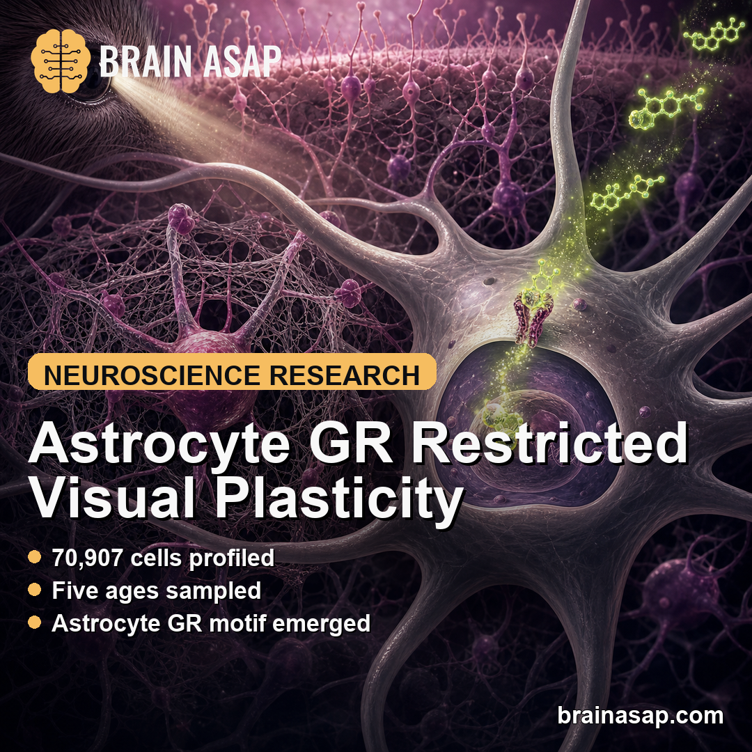
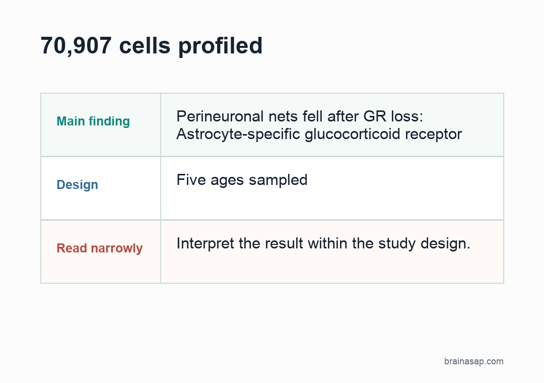
- 70,907 cells profiled: SHARE-seq mapped RNA expression and chromatin accessibility across visual cortex development in normal-reared and dark-reared mice.
- Five ages sampled: Researchers examined postnatal days 7, 14, 21, 28, and 35, spanning eye opening, the ocular dominance critical period, and critical period closure.
- Astrocyte GR motif emerged: Glucocorticoid receptor and mineralocorticoid receptor motifs were among the top experience-induced regulatory patterns in astrocytes.
- Perineuronal nets fell after GR loss: Astrocyte-specific glucocorticoid receptor deletion reduced perineuronal net density on parvalbumin interneurons in visual cortex.
- Adult plasticity reopened: Removing astrocyte glucocorticoid receptor signaling in mature visual cortex enabled ocular dominance plasticity after 3-4 days of monocular deprivation.
Source: Nature (2026) | Gegenhuber et al.
Astrocytes are often described as support cells, but this study treated them as active regulators of when cortical circuits stop being highly plastic. The focus was the mouse primary visual cortex, a standard system for studying critical periods in brain development.
During a critical period, visual experience can strongly reshape cortical responses. Later in development, the same short deprivation usually has a weaker effect because the circuit has matured and structural brakes have formed.
Visual Experience Changed Gene Programs Across the Developing Cortex
Researchers used SHARE-seq, a single-cell method that pairs RNA expression with chromatin accessibility, to profile visual cortex from normal-reared and dark-reared mice. The dataset covered 70,907 cells across excitatory neurons, inhibitory neurons, astrocytes, oligodendrocytes, microglia, endothelial cells, and related cortical populations.
The developmental sampling was built around visual milestones. The team profiled P7 before eye opening, P14 around eye opening, P21-P28 during the ocular dominance critical period, and P35 near critical period closure.
Light exposure produced gene-expression and chromatin changes across cortical cell types. About 40% of gene changes occurred in multiple cell types and included immediate-early genes such as Fosl2, Egr1, and Nr4a1.
Other changes were cell-type-specific. Those included genes involved in synaptic wiring, cell-surface communication, secreted proteins, and maturation programs that differed between neuronal and non-neuronal cells.
- Neuronal response: Excitatory and inhibitory neurons showed strong experience-linked programs around P21 and P35.
- Astrocyte response: Astrocytes showed a distinct regulatory pattern involving nuclear hormone receptor motifs.
- Validation step: MERFISH with a 500-gene panel confirmed several experience-responsive genes in visual cortex tissue.
Astrocyte Glucocorticoid Receptor Signaling Marked Circuit Maturation
In astrocytes, the top experience-induced regulatory pattern differed from the neuronal pattern. Motifs for the glucocorticoid receptor, encoded by Nr3c1, and related nuclear receptor factors emerged as major astrocyte-specific marks.
Glucocorticoid receptor activity is one route by which circulating stress hormones can alter gene regulation. The receptor is present before activation, then moves into the nucleus after hormone binding and changes transcription at target sites.
The study connected that hormone-responsive system to astrocyte maturation. Light exposure recruited glucocorticoid receptor to astrocyte chromatin, and the resulting gene program partly overlapped with human brain development patterns.
The astrocyte program affected extracellular matrix genes. That detail is important because extracellular matrix organization helps stabilize cortical circuits and reduce plasticity after developmental windows close.
Deleting Astrocyte GR Reduced Perineuronal Nets on PV Interneurons
The team then tested function by deleting glucocorticoid receptor in astrocytes. Loss of astrocyte GR shifted extracellular matrix gene expression toward a less mature profile, including higher immature glycoprotein and matrix-degrading enzyme cues.
Researchers measured perineuronal nets, extracellular matrix structures that form around parvalbumin-positive inhibitory neurons and help restrain ocular dominance plasticity. Astrocyte GR deletion reduced perineuronal net density across visual cortex layers at P35, with strong effects in layers 4 and 5.
That was not only a developmental finding. Deleting astrocyte GR in adult visual cortex also reduced perineuronal net and aggrecan levels, suggesting the pathway helps maintain mature circuit brakes after the early critical period.
The intervention separated several related circuit features:
- Perineuronal nets: Wisteria floribunda lectin signal on parvalbumin cell bodies decreased after astrocyte GR deletion.
- PV expression: Parvalbumin expression was reduced, but the number of PV interneurons was not the main changed measure.
- Corticosterone link: Systemic corticosterone around eye opening increased perineuronal net density in dark-reared mice.
Adult Astrocyte GR Loss Reopened Ocular Dominance Plasticity
The strongest circuit test used adult mice. Researchers delivered astrocyte-selective viral tools to remove GR in mature visual cortex, then deprived one eye for 3-4 days and recorded responses from binocular visual cortex.
Normally, that short deprivation is not enough to produce ocular dominance plasticity in adult mice. After astrocyte GR loss, mature circuits responded: the open eye gained relative influence and the deprived eye response weakened.
This experiment supports a specific claim. Astrocyte glucocorticoid receptor activity is not only correlated with development; removing it in adult visual cortex was sufficient to re-engage a plasticity response after brief monocular deprivation.
The pathway also gives early-life stress biology a mechanistic foothold. Glucocorticoids are stress hormones, and abnormal critical-period timing has been discussed in neurodevelopmental disorders.
The experiment did not model autism or schizophrenia directly, but it identified a brain-cell route by which hormone exposure could alter maturation timing.
The Finding Is Mechanistic, Not a Call to Block Stress Hormones
The evidence places astrocytes in the plasticity brake for visual cortex through glucocorticoid receptor-driven maturation programs. It also places astrocytes between sensory experience, hormone exposure, extracellular matrix formation, and circuit closure.
Several limits keep the claim narrow. The work was done in mice, the circuit was visual cortex, and the interventions were targeted experimental manipulations, not drug treatments for people.
The next clinical question is not whether stress hormones are good or bad in general. The sharper question is how timing, cell type, dose, and developmental stage shape hormone effects on astrocytes and circuit plasticity.
Citation: DOI: 10.1038/s41586-026-10512-9. Gegenhuber et al. Astrocyte glucocorticoid receptor signalling restricts neuronal plasticity. Nature. 2026.
Study Design: Mouse visual cortex development study combining single-cell multiomics, MERFISH validation, astrocyte-specific glucocorticoid receptor deletion, perineuronal net assays, and ocular dominance physiology.
Sample/Model: Normal-reared and dark-reared mice across postnatal development, with 70,907 visual cortex cells profiled by SHARE-seq.
Key Statistic: Adult astrocyte GR deletion enabled ocular dominance plasticity after 3-4 days of monocular deprivation, a short deprivation window that normally does not produce this adult response.
Caveat: The study identifies a mouse visual cortex mechanism and does not test a human therapy or a generalized stress-hormone intervention.